
|
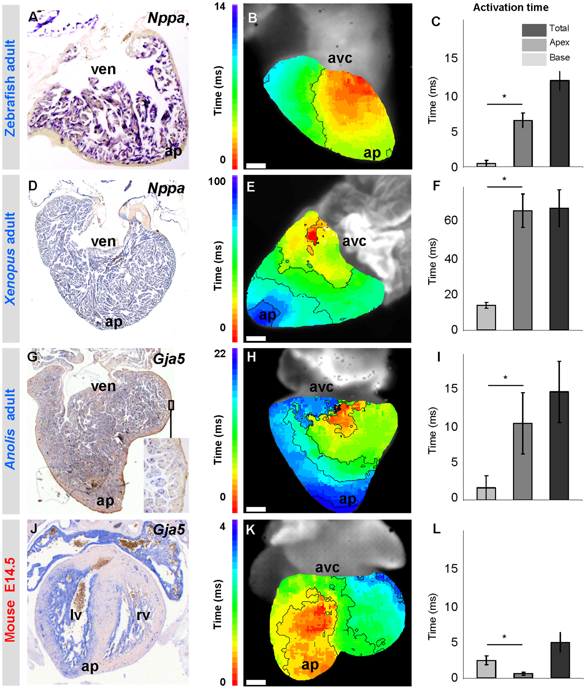
Fig. 9
Trabeculated ventricles are activated from base to apex.
(A, D, G, J) Markers of fast propagating myocardium (Nppa and Gja5) are homogenously expressed in the ventricular trabeculated myocardium from base to apex (ap). (B, E, H) Ventricular activation occurs from base to apex. Early activation is red, late activation is blue. Note that the time-colour coding in panel E is different from that in panels B and H. (K) In species with thick compact myocardium, surface breakthrough of the activation front is earlier in the apical region than in the base. (C, F, I, L) Graphs show the average activation time of the apex and base and the total ventricular activation time. Note that in zebrafish, Xenopus and Anolis, the ventricular base is activated earlier than the apex whereas in mice the ventricular base is activated later than the apex (* Significantly different (one-way ANOVA P<0.05)). n is 3, 6, 9 and 2, respectively. Scale bars in (B, E, H, K) indicate respectively 0.2, 1, 0.5 and 0.1 millimetre. avc, atrioventricular canal; ven, ventricle.