
- Title
-
Novel expression patterns of metabotropic glutamate receptor 6 in the zebrafish nervous system
- Authors
- Huang, Y.Y., Haug, M.F., Gesemann, M., and Neuhauss, S.C.
- Source
- Full text @ PLoS One

RNA expression of zebrafish mglur6 and gnao paralogs. RNA expression of mglur6a (A1–A4), mglur6b (B1–B4), gnaoa (C1–C4) and gnaob (D1–D4) in dorsal (1) and lateral (2) views and in isolated eyes (3) of a 5 dpf zebrafish, as well as in adult retinal cross-sections (4). A1, A2: Expression of mglur6a is visible in the habenula (Ha), the medial (TeO) and lateral (lTeO) tectum opticum, the midbrain (mb), a part of the mid-hindbrain boundary (mhb) and a bilateral nucleus of the medulla oblongata (MO). A3: In an eye separated from a whole mount stained larva mglur6a is expressed in the proximal inner nuclear layer (INL, arrow) and the ganglion cell layer (GCL). A4: Additional to the cellular expression in the proximal INL (arrows) and the GCL, mglur6a labels cells in the medial INL (arrowheads) in adult. B1, B2: mglur6b reveals a staining in the olfactory bulb (OB) and a weak labeling of a part of the diencephalon (di). B3: The isolated eye shows, mglur6b expression in the medial INL (arrowhead), the proximal INL (arrow) and the GCL. B4: The adult retinal cross section shows the same localization for mglur6b as the larval fish, however, the staining in the medial INL is restricted to a subset of cells (arrowheads). C1, C2: gnaoa is expressed in the olfactory bulb (OB), the pallium (P), the habenula (Ha), the tectum opticum (TeO) and in all cranial ganglia (CG, arrowheads). C3: The retina reveals gnaoa labeling in the proximal INL (arrow) and the GCL. C4: Similar to the larval retina, gnaoa labels the proximal INL (arrow) and the GCL in the adult retina. Additionally, gnaoa stains weakly a subset of cells in the medial INL (arrowheads). D1, D2: gnaob shows no expression in the brain. D3: The expression of gnaob in the larval fish eye is restricted to the medial INL (arrow) and the GCL. D4: Similar to the larval eye, in adult retinal cross-sections gnaob is located in cells of the medial INL and the GCL. All scale bars = 40 μm. Scale bar in A1 applies to all whole mount images, scale bar in A3 applies to A3, B3, C3 and D3, scale bar in A4 applies to A4, B4, C4 and D4. EXPRESSION / LABELING:
|
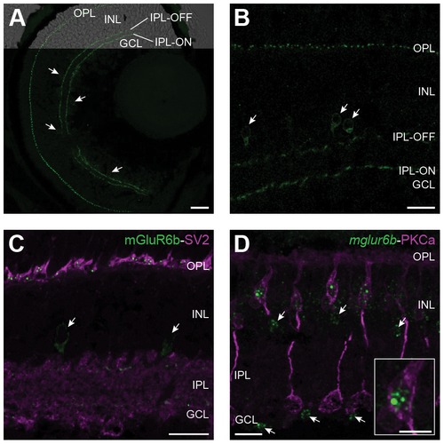
Subcellular localization of mGluR6b in the zebrafish retina. Z-projections of confocal image stacks of immunohistochemically labeled larval and adult retinal cross-sections. A: A larval retina at 5 dpf stained with the mGluR6b antibody shows labeling in the outer plexiform layer (OPL) and in an ON- and an OFF-layer of the inner plexiform layer (IPL). In addition, single cells adjacent to the IPL in the INL and the GCL (arrowheads) express mGluR6b as well. Scale bar = 20 μm. B: In the adult zebrafish retina, mGluR6b stains similar structures as in the larval retina with the exception that we could only detect labeled cells in the inner part of the INL (arrowhead) but not in the GCL. Scale bar = 15 μm. C: A doublelabeling of mGluR6b (green) and SV2 (magenta) in an adult retinal cross-section reveals the postsynaptic localization of mGluR6b in the OPL. Again, an mGluR6b expression in single cells of the proximal IPL is detected (arrows). Scale bar = 15 μm. D: Fluorescent in situ hybridization of mglur6b (green) combined with an immunohistochemical labeling of ON-bipolar cells by PKCalpha (magenta) confirms the localization of mGluR6b in ON-bipolar cells of the INL. Scale bar = 15 μm. Arrows point to cells of the proximal INL and the GCL expressing mglur6b. The insert shows a close up of an ON-bipolar cell body and its fluorescent mglur6b signal in the cytosol. Scale bar = 5 μm. EXPRESSION / LABELING:
|

Electroretinogram recordings of mglur6b-depleted zebrafish larvae. The downregulation of mGluR6b leads to a dose dependent decrease of the ERG b-wave in 5 day old zebrafish larvae indicating a diminished ON-response. A: Plotted b-wave amplitudes at different light intensities. Control Morpholino (MO) injected larvae of different concentrations showed no significant differences in b-wave amplitude, neither among each other nor in comparison to uninjected wild type larvae (data not shown). Therefore, recordings of all control MO larvae (n = 46) were taken together to build one curve. Injection of mglur6b MO leads to a dose dependent depletion of the b-wave (1.5 ng mglur6b MO: n = 14; 2.99 ng MO: n = 24; 7.48 ng MO: n = 13). All data points represent the means ± SEM. B: Significance of the b-wave amplitude reduction was calculated using a 2-way ANOVA (* = p<0.1; ** = p<0.01; *** = p<0.001). PHENOTYPE:
|

mGluR6b expression in the mglur6b-depleted zebrafish retina at 5 dpf. Immunohistochemical analysis using the mGluR6b antibody confirms the downregulation of mGluR6b in the 5 dpf zebrafish retina. A: mGluR6b expression in a non-injected sibling. B: Injection of 2.5 ng mglur6b MO leads to an incomplete downregulation of the mGluR6b protein since a faint staining in the plexiform layers is still visible. C: 6.7 ng mglur6b antisense-RNA lead to a complete knockdown of the mGluR6b protein in 5 dpf zebrafish retinas. Scale bar (applies for all images A–C) = 20 μm. |

Cross-absorbance of the mGluR6b antibody. A: Immunohistochemistry image of a cross section through a 5 day old larval retina stained with the original mGluR6b antibody (1:200). B: Confocal image of a larval retina at 5 dpf stained with the cross-absorbed mGluR6b antibody (1:150). For further description see Figure 3. Scale bar in B = 20 μm (applies for A and B). C: Dot-blot analysis showing the increased specificity of the cross-absorbed mGluR6b antibody. 1 µg of mGluR6a (6a) and mGluR6b (6b) epitopes were pipetted on nitrocellulose membranes (0.45 μm; Bio-Rad, Reinach, Switzerland) and incubated with the non cross-absorbed and the cross-absorbed antibodies. Following cross-absorbing the epitope of the mGluR6a is not recognized anymore. |