
- Title
-
Dynamic expression patterns of 6-O endosulfatases during zebrafish development suggest a subfunctionalisation event for sulf2
- Authors
- Gorsi, B., Whelan, S., and Stringer, S.E.
- Source
- Full text @ Dev. Dyn.
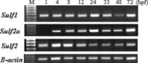
RT-PCR of sulf transcripts during zebrafish development. Sulf1,Sulf2a, and Sulf2 primers were designed to amplify 512, 498, and 687 bp, respectively, with inclusion of the ATG start site. β-actin primers were designed to amplify 298 bp. The resulting PCR products were electrophoresed on a 1% agarose gel. The intensity of a 300-bp DNA fragment amplified using β-actin-specific primers was used to evaluate the relative amount of cDNA used in each PCR. Marker lane represents 1-kb ladder. EXPRESSION / LABELING:
|
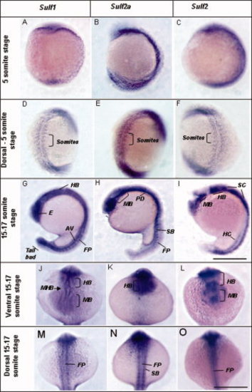
Expression of heparan sulfate (HS) sulf genes during early zebrafish development. Expression of sulf1 at 5-somite stage, lateral view (A) and dorsal view (D) and 15–17-somite stage, lateral (G), ventral (J), and dorsal views (M). Expression of sulf2a at 5-somite stage, lateral view (B) and dorsal view (E), and 15–17-somite stage, lateral (H), ventral (K), and dorsal view (N), Expression of sulf2 at 5-somite stage, lateral view (C) and dorsal view (F), and 15–17-somite stage, lateral (I), ventral (L), and dorsal view (O). AV, axial vessels; E, eye; FP, floorplate; HB, hindbrain; HC, hypochord; MB, midbrain; PD, pronephric ducts; SB, somite boundaries. SC, spinal cord. Scale bar = 200 μm. EXPRESSION / LABELING:
|
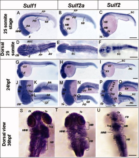
Expression of sulfs during segmentation and pharyngula period. Expression of sulf1 at 25-somite stage, lateral (A) and dorsal view (D); lateral view at 24hpf (G); close up of head, anterior trunk, and tail (J–L); and 36hpf dorsal view (S). Expression of sulf2a at 25-somite stage, lateral (B) and dorsal view (E); lateral view at 24hpf (H); close up of head, anterior trunk, and tail (M–O); and 36hpf dorsal view (T). Expression of sulf2 at 25-somite stage, lateral (C) and dorsal view (F); lateral view at 24hpf (I); close up of head, anterior trunk, and tail (P–R); and 36hpf dorsal view (U). ASC, anterior spinal cord; AV, axial vessels; BA, branchial arches; Cb, cerebellum; CP; caudal plexus; Di, diencephalon; E, eye; Ep, epiphysis; FB, forebrain; FP, floorplate; HB, hindbrain; HC, hypochord; M, mesencephalon; MHB, mid-hindbrain; OB, olfactory bulb; OT, otic vesicle; PD, pronephric ducts; R, rhombomeres; SB, somite boundaries; SC, spinal cord; SN; spinal cord neurons; Te, telencephalon; Teg, tegmentum. Scale bar = 200 μm. EXPRESSION / LABELING:
|
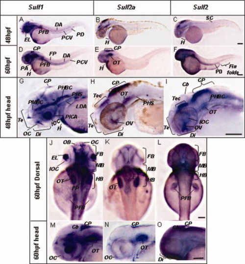
Expression of sulfs during the long-pec and pec fin stage. Expression of sulf1 at 48hpf whole shot; arrow denotes caudal fin folds (A) and lateral view of head (G), whole shot of embryo at 60hpf (D), lateral head (M), and dorsal view (J). Expression of sulf2a at 48hpf whole shot (B) and lateral view of head (H), whole shot of embryo at 60hpf (E), lateral head (N), and dorsal view (K). Expression of sulf2 at 48hpf whole shot (C) and lateral view of head (I), whole shot of embryo at 60hpf (F), lateral head (O), and dorsal view (L). Cb, cerebellum; CP; choroid plexus; DA, dorsal aorta; Di, diencephalon; E, eye; EL, eye lens; FB, forebrain; FP, floorplate; H, heart; HB, hindbrain; IOC, inner optic circle; LDA, lateral dorsal aorta; MB, midbrain; OB, olfactory bulb; OC, oral cavity; OT, otic vesicle; PA, pharyngeal arch; PCV, posterior cardinal vein; PD, pronephric ducts; PFB, pectoral finbuds; PHBC, primordial hindbrain channel; PHS, primary head sinus; SB, somite boundaries; SC, spinal cord; Te, telencephalon; Teg, tegmentum. Scale bar = 200 μm. EXPRESSION / LABELING:
|
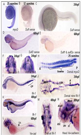
Expression of sulf sense probes and endothelial and muscle markers during zebrafish development. Representative images of sulf sense probes at 17 somite stage (B), 24hpf (C)and 48hpf (D,E). Markers used as positive control for all WISH experiments, myoD expression at 17 somite stage (A) dorsal ciew of somite staining(H). Expression pan-endothelial marker flk-1 at 24hpf lateral view of whole embryos (I) and dorsal view of flat mount of (J), dorsal (F) and ventral view (G) of head at 24hpf. Lateral view of flk-1 expression at 48hpf (K) and dorsal view (M). Lateral view of embryo expressing ve-cad at 60hpf (L) and close up of the head of embryo (N). AA, aortic arches; ACeV, anterior cerebral vein; AV, axial vessels; CP; caudal plexus; E, eye; EL, eye lens; H, heart; IOC, inner optic circle; LDA, lateral dorsal aortae; MCeV middle cerebral vein; OV, optic vein; PHBC, primordial hindbrain channel; PICA, primitive internal cartoid artery; PrA, prosencephalic artery. |