
- Title
-
The expression patterns of minor fibrillar collagens during development in zebrafish
- Authors
- Fang, M., Adams, J.S., McMahan, B.L., Brown, R.J., and Oxford, J.T.
- Source
- Full text @ Gene Expr. Patterns

Temporal expression and splicing patterns of minor fibrillar collagens. RT-PCR was performed using RNA extracted from wild type embryos at 4, 6, 10, 16, 24, 36, 48, 72 hpf. (A) Multiple bands were observed when cDNA was amplified within the region between exons 5 and 9 in the case of Col11a1a on chr24, Col11a2 on chr19, and Col5a3 on chr3. Single amplification products were observed in the case of Col5a1 and Col11a1b on chr2. (B) Splicing patterns are depicted, reflecting the exon usage for those genes undergoing alternative splicing within the variable region. Boxes represent individual exons within the VR, whereas lines in between represent introns. Relative size of all elements depicted reflects relative actual size in basepairs. For Col11a1a on chr24, the predominant spliceform at the earliest stages examined in this study included exons 6A, 7 and 8 within the variable region. Other minor transcripts observed included exons 6A and 6B or 7 alone. For Col11a2, exons 6, 7 or 8 were observed to be either included or excluded in a complex pattern. An additional exon of approximately 1.2 kb was observed, replacing exons 6, 7 and 8 in the case of the largest alternatively spliced form. In the case of Col5a3, alternative splicing involved exon 7. (C). Each amplification product was excised and sequenced to confirm identity, intron–exon boundaries, and exon usage in comparison to genomic sequence currently available. Variable region exon sequences in C are color-coded to match the block diagram in (B). EXPRESSION / LABELING:
|

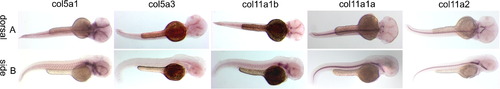
Spatial expression patterns of minor fibrillar collagen genes in zebrafish at 16 hpf by whole mount in situ hybridization. The expression patterns of Col5a1 (A, F and K), Col5a3 (B and G), Col11a1b (C, H and L), Col11a1a (D, I and M) and Col11a2 (E and J) are shown in both dorsal (A–E) and lateral (F–J) views. Arrows in (D) and (H) indicate expression in the hindbrain. Magnified views of (A), (H), and (D) demonstrate Col5a1 expression in neural crest (K), Col11a1b (L) and Col11a1a (M) in hindbrain (arrow in (L) and lower arrow in (M)) and otic placode (upper arrow in (M)). EXPRESSION / LABELING:
|
Spatial expression patterns of minor fibrillar collagen genes in zebrafish at 24 hpf by whole mount in situ hybridization. The expression patterns of Col5a1, Col5a3, Col11a1b, Col11a1a and Col11a2 are shown in both dorsal (A) and lateral (B) views. Magnified views demonstrate expression in notochord and region of developing somites (C). Magnified view in column 5, row C depicts Col11a2 expression in the developing eye. EXPRESSION / LABELING:
|
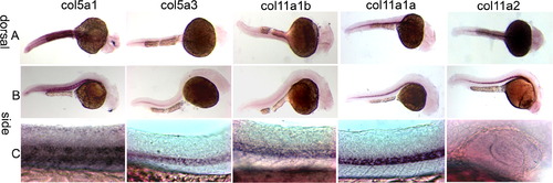
Spatial expression patterns of minor fibrillar collagen genes in zebrafish at 48 hpf by whole mount in situ hybridization. The expression patterns of Col5a1, Col5a3, Col11a1b, Col11a1a and Col11a2 are shown in both dorsal (A) and lateral (B) views. Expression is observed in the perioptic mesoderm, developing craniofacial cartilages, and presumptive retinal pigmented epithelium. Higher magnification views of patterns of expression are shown in Fig. 6 for 72 hpf. EXPRESSION / LABELING:
|
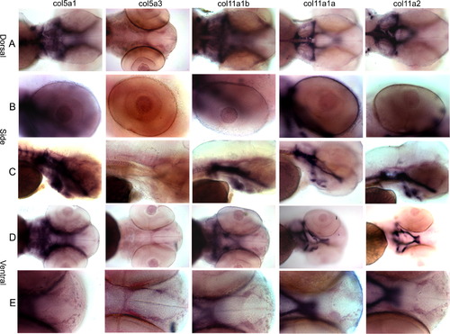
Spatial expression patterns of minor fibrillar collagen genes in zebrafish at 72 hpf by whole mount in situ hybridization. The expression patterns of Col5a1, Col5a3, Col11a1b, Col11a1a and Col11a2 are shown in dorsal (A), lateral (B and C), and ventral (D and E) views. A comparison of overall craniofacial staining is shown in (A), (C) and (D), demonstrating staining in the cranial cartilages including the trabeculae, ceratohyal, ethmoid plate, Meckel’s cartilage, polar cartilage, palatoquadrate and parachordal cartilage. Expression in the eye is shown in row (B). Anterior-most view of developing craniofacial region is shown in row (E). EXPRESSION / LABELING:
|

Expression of minor fibrillar collagens at 0 hpf in zebrafish. Only Col11a2 shows maternal transcription. EXPRESSION / LABELING:
|
Reprinted from Gene expression patterns : GEP, 10(7-8), Fang, M., Adams, J.S., McMahan, B.L., Brown, R.J., and Oxford, J.T., The expression patterns of minor fibrillar collagens during development in zebrafish, 315-322, Copyright (2010) with permission from Elsevier. Full text @ Gene Expr. Patterns