
Fig. 6
|
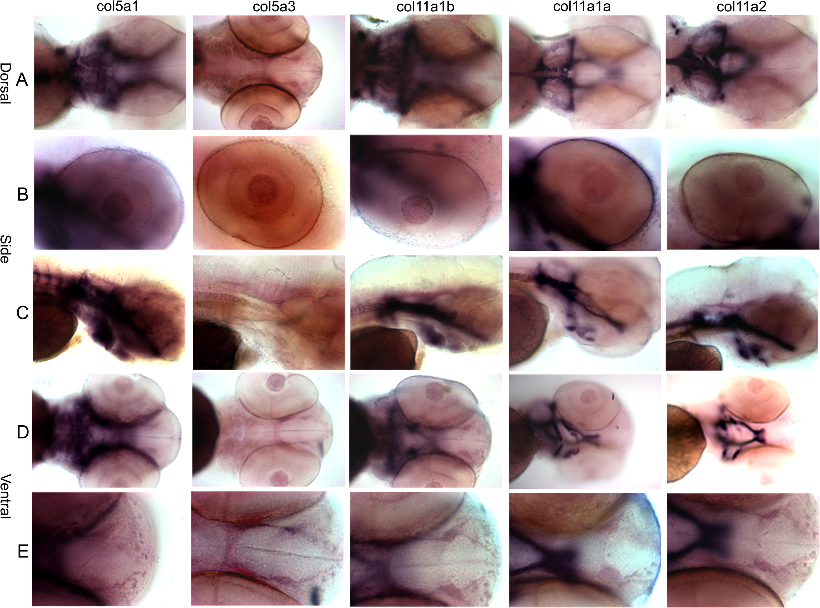
Fig. 6 Spatial expression patterns of minor fibrillar collagen genes in zebrafish at 72 hpf by whole mount in situ hybridization. The expression patterns of Col5a1, Col5a3, Col11a1b, Col11a1a and Col11a2 are shown in dorsal (A), lateral (B and C), and ventral (D and E) views. A comparison of overall craniofacial staining is shown in (A), (C) and (D), demonstrating staining in the cranial cartilages including the trabeculae, ceratohyal, ethmoid plate, Meckel’s cartilage, polar cartilage, palatoquadrate and parachordal cartilage. Expression in the eye is shown in row (B). Anterior-most view of developing craniofacial region is shown in row (E).
Reprinted from Gene expression patterns : GEP, 10(7-8), Fang, M., Adams, J.S., McMahan, B.L., Brown, R.J., and Oxford, J.T., The expression patterns of minor fibrillar collagens during development in zebrafish, 315-322, Copyright (2010) with permission from Elsevier. Full text @ Gene Expr. Patterns