
- Title
-
Cloning of hif-1alpha and hif-2alpha and mRNA expression pattern during development in zebrafish
- Authors
- Rojas, D.A., Perez-Munizaga, D.A., Centanin, L., Antonelli, M., Wappner, P., Allende, M.L., and Reyes, A.E.
- Source
- Full text @ Gene Expr. Patterns
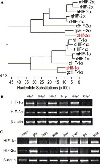
Phylogenetic tree and expression of hif-1α and hif-2α during embryonic development. (A) Phylogenetic tree resulting from CLUSTAL analysis of HIF protein sequences. Horizontal lines indicate the degree of relatedness. Sequences are from human (h), mouse (m), rat (r), quail (q), chicken (c), Xenopus tropicalis (xt), grass carp (gc), dog (d), squirrel (s) and zebrafish (z). (B) Expression of hif-1α and hif-2α at different stages of development as assayed by RT-PCR from total RNA prepared from the indicated stages. hif-1α and hif-2α are expressed in all the stages analyzed. (C) Expression of hif-1α and hif-2α in adult tissues. Total RNA was prepared from the indicated tissues from adult fishes (one year old); both genes are expressed in all adult tissues analyzed, although at variable levels. As an internal control, expression of β-actin was assessed in the same samples (lower panel in B and C). |
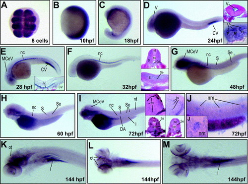
Whole-mount in situ hybridization for hif-1α. Expression of hif-1α mRNA was analyzed at the indicated developmental stages. hif-1α mRNA is detected at early stages in an ubiquitous expression pattern (A and B), becoming ventrally restricted by late embryogenesis (C); A, dorsal view; B and C, lateral view with anterior on top. At 24 hpf hif-1α is expressed in ventricle epithelia (D, paraffin transverse cross-section in top inset), as confirmed by Nomarski microscopy (D, bottom inset). At 28 hpf, expression is present in the notochord, caudal vein and brain blood vessels (E), as confirmed by histological sagittal cross-section (inset in E). At 32 hpf, hif-1α mRNA is detected in brain and notochord (F), paraffin cross-section of embryos shows that expression occurs in the somites, notochord and dorsal aorta (transverse cross-section, top inset in F) as well as in intersegmental blood vessels (sagittal cross-section, bottom inset in F). At 48 and 60 hpf (G and H, respectively) the expression of hif-1α is detected in the brain, notochord, somites and intersegmental blood vessels. At 72 hpf the mRNA is detected in the brain, branchial region, intersegmental blood vessels, dorsal aorta and intestine (I), left insets in I show a Tg (fli1:EGFP)y1 embryo subjected to GFP immunohistochemistry and hif-1α mRNA in situ hybridization; right insets in I show the mRNA in situ hybridization pattern of hif-1α in a wild type fish (all insets are transverse cross-sections). Overstaining of 72 hpf embryos revealed expression in caudal neuromasts (J), as confirmed by DIC optics (inset in J). At 144 hpf expression was observed in the optic tectum, retina and intestine (K). A dorsal view of embryos at 144 hpf shows expression in the optic tectum (L) and retina (M). In (D) to (M) lateral view, with anterior to the left. Insets showing transverse cross-sections with dorsal side up; sagittal cross-sections show the anterior to the left. Abbreviations: V, ventricle epithelia; F, forebrain; M, midbrain; H, hindbrain; MCeV, middle cerebral vein; CV, caudal vein; Se, intersegmental vessel; DA, dorsal aorta; nc, notochord; S, somites; nt, neural tube; I, intestine; bv, blood vessel; nm, neuromasts; ot, optic tectum; e, eye; r, retina. |
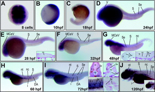
Expression of hif-2α in zebrafish embryos and larvae as revealed by mRNA in situ hybridization. hif-2α mRNA is maternally expressed and ubiquitously distributed at early stages (A and B), becoming ventrally restricted later in embryogenesis (C); (A) dorsal view; (B) and (C), lateral views with the anterior side on top. At 24 (D), 28 (E) and 32 hpf (F) hif-2α was detected in the brain, somites, and blood vessels. At 48 hpf, hif-2α expression is present in brain blood vessels, notochord and intersegmental blood vessels (G), as it is shown in the brain (left inset in G) and tail paraffin cross-sections (right inset in G). At 60 hpf, hif-2α mRNA is expressed in the brain, somites, dorsal aorta, intestine and notochord (H). At 72 hpf, hif-2α expression was detected in the brain, brain blood vessels, intersegmental vessels, dorsal aorta and intestine (I). mRNA in situ hybridization for hif-2α and anti-GFP immunohistochemistry in Tg(fli1:EGFP)y1 embryos show that expression occurs in intersegmental blood vessels (left-top inset in I, transverse cross-section; left-bottom inset, DIC lateral view). Paraffin cross-sections of wild type embryos at 72 hpf, show expression of hif-2α in somites, intersegmental blood vessels, notochord and aorta (right-top inset in I, transverse cross-section; right-bottom in I, sagittal cross-section). At 120 hpf, expression of hif-2α was detected in the optic tectum, intersegmental blood vessels, notochord, intestine and retina (J). (D)–(J) are lateral views with anterior to the left. Insets show transverse cross-sections with the dorsal part on the top, or sagittal cross-sections with anterior to the left. Abbreviations: V, ventricle epithelia; F, forebrain; M, midbrain; H, hindbrain; MCeV, middle cerebral vein; Se, intersegmental vessel; DA, dorsal aorta; nc, notochord; S, somites; nt, neural tube; I, intestine; bv, blood vessel; nm, neuromasts; ot, optic tectum; e, eye; r, retina. |
Reprinted from Gene expression patterns : GEP, 7(3), Rojas, D.A., Perez-Munizaga, D.A., Centanin, L., Antonelli, M., Wappner, P., Allende, M.L., and Reyes, A.E., Cloning of hif-1alpha and hif-2alpha and mRNA expression pattern during development in zebrafish, 339-345, Copyright (2007) with permission from Elsevier. Full text @ Gene Expr. Patterns