
- Title
-
CFTR deficiency causes cardiac dysplasia during zebrafish embryogenesis and is associated with dilated cardiomyopathy
- Authors
- Liu, Y., Lin, Z., Liu, M., Liao, H., Chen, Y., Zhang, X., Chan, H.C., Zhou, B., Rao, L., Sun, H.
- Source
- Full text @ Mech. Dev.

cftr mutation induces cardiac dysplasia in the early zebrafish embryo. (A) Lateral views of live zebrafish embryos at 60 hpf. Arrows indicate the pericardium. Heart rate (B) and quantification of ventricular fractional shortening (FS) (C) are represented in the scatter diagram. (D) The expression pattern of nkx2.5 at 13 hpf is significantly affected in cftr mutant embryos. Embryo orientations: dorsal view with anterior oriented at the top. Arrows indicate normal nkx2.5 expression; arrowheads demonstrate the aberrant strength and position of nkx2.5 expression in the cftr mutant; double-ended arrows indicate the distance between the two populations expressing nkx2.5. Relative expression of nkx2.5 (E) and relative distance of nkx2.5 lateral signal (F) are represented in the scatter diagram. (G) Heart morphology (marked by the pan-cardiomyocyte marker myl7) at 48 hpf was impaired significantly in cftr mutants. Embryo orientation: ventral view with the anterior at the top. Arrows point to the atrium; red lines demonstrate the acute angle formed between the atrial and ventricular axes. Looping angle (H) and area ratio of atrium/ventricle (I) are represented in the scatter diagram. The expression of bmp4 (J and K) and has2 (L and M) is dramatically reduced in cftr mutant embryos at 48 hpf. Embryo orientation: ventral views with the anterior at the top. Arrows point to the signal generated by detected marker genes. EXPRESSION / LABELING:
PHENOTYPE:
|

Treatment with the CFTR inhibitor CFTR_inh172 at the blastula stage leads to cardiac dysplasia in early zebrafish embryos. (A–C) The expression pattern of nkx2.5 at 13 hpf is significantly affected. Embryo orientations: dorsal view with anterior oriented at the top. Arrows indicate normal nkx2.5 expression; arrowheads demonstrate the aberrant strength and position of nkx2.5 expression in the cftr mutant; double-ended arrows indicate the distance between the two populations expressing nkx2.5. (D and E) Heart morphology (marked by the pan-cardiomyocyte marker myl7) at 24 hpf was altered significantly. Embryo orientation: ventral view with the anterior at the top. Red lines demonstrate the acute angle formed between the head midline and heart tube. (F–H) Heart morphology (marked by myl7) at 48 hpf was impaired significantly. Embryo orientation: ventral view with the anterior at the top. Arrows point to the atrium; red lines demonstrate the acute angle formed between the atrial and ventricular axes. EXPRESSION / LABELING:
PHENOTYPE:
|

ZFIN is incorporating published figure images and captions as part of an ongoing project. Figures from some publications have not yet been curated, or are not available for display because of copyright restrictions. |
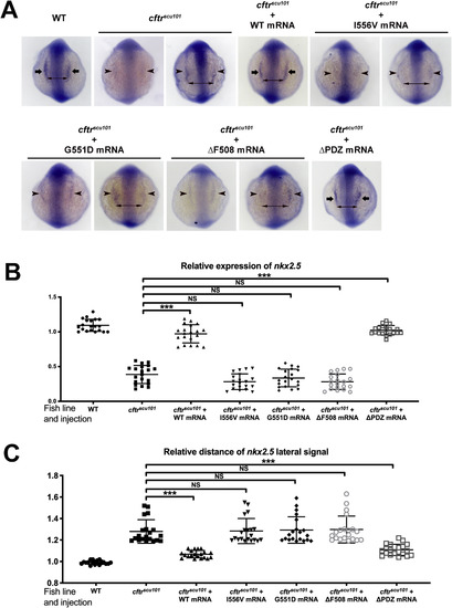
Fig. 4. CFTR I556V mutants fail to rescue nkx2.5-expressing cardiac progenitor cells at the 8-somite stage (13 hpf) of embryogenesis. (A) Embryo orientations: dorsal view with anterior at the top. Arrows point to the normal nkx2.5 signal; arrowheads demonstrate the aberrant strength and position of nkx2.5 expression induced by cftr mutants; double-ended arrows indicate the distance between the two nkx2.5+ progenitor populations. Relative expression of nkx2.5 (B) and relative distance of nkx2.5 lateral signal (C) are represented in the scatter diagram. EXPRESSION / LABELING:
PHENOTYPE:
|
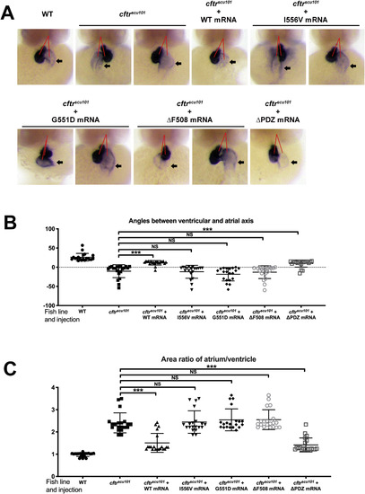
CFTR I556V fail to rescue heart morphology marked by myl7 expression 48 hpf during embryogenesis. (A) Embryo orientations: ventral views with the anterior at the top. Arrows point to the atrium; red lines indicate the acute angle formed between the atrial and ventricular axes. Looping angle (B) and area ratio of atrium/ventricle (C) are represented in the scatter diagram. EXPRESSION / LABELING:
|
Reprinted from Mechanisms of Development, 163, Liu, Y., Lin, Z., Liu, M., Liao, H., Chen, Y., Zhang, X., Chan, H.C., Zhou, B., Rao, L., Sun, H., CFTR deficiency causes cardiac dysplasia during zebrafish embryogenesis and is associated with dilated cardiomyopathy, 103627, Copyright (2020) with permission from Elsevier. Full text @ Mech. Dev.