
- Title
-
Identification of target genes downstream of Semaphorin6A/PlexinA2 signaling in zebrafish
- Authors
- Emerson, S.E., St Clair, R.M., Waldron, A.L., Bruno, S.R., Duong, A., Driscoll, H.E., Ballif, B.A., McFarlane, S., Ebert, A.M.
- Source
- Full text @ Dev. Dyn.
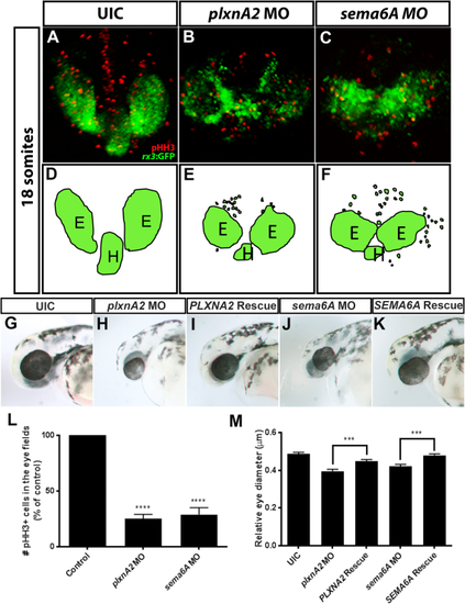
Sema6A and plxnA2 knockdown leads to loss of cohesion and decreased proliferation in eye vesicles. A–C: Confocal dorsal views of 18 somite stage rx3:GFP embryos co-labeled with pHH3. L: Both sema6A and plxnA2 morphants display ectopic retinal precursor cells outside of the eye field as well as significantly reduced pHH3 positive cells in the developing eyes compared with controls, as quantified (control 100%, N = 3, n = 30; plxnA2 MO 25.11% ± 0.72%, N = 3, n = 30, ****P < 0.0001; sema6A MO 28.86% ± 1.12%, N = 3, n = 30; ****P < 0.0001, one-way ANOVA, multiple comparisons post-hoc test). D–F: Schematics of rx3:GFP dorsal view. G,H,J,M: Decreased proliferation is later observed as smaller eyes at 48 hpf (control 0.49 μm ± 0.008 μm, N = 3, n = 30; plxnA2 MO 0.40 μm ± 0.01 μm, N = 3, n = 29, ****P < 0.0001; sema6A MO 0.42 μm ± 0.009 μm, N = 3, n = 31; ****P < 0.0001, one-way ANOVA, multiple comparisons test). G,I,K,M: Full length human PLXNA2 and SEMA6A mRNA partially rescues smaller eye phenotypes (control 0.49 μm ± 0.008 μm, N = 3, n = 30; PLXNA2 rescue 0.45 μm ± 0.009 μm, N = 3, n = 32, ***P < 0.001; SEMA6A rescue 0.48 μm ± 0.008 μm, N = 3, n = 28; ***P < 0.001, one-way ANOVA, multiple comparisons test. Error bars indicate SEM). E, eye; H, hypothalamus. EXPRESSION / LABELING:
PHENOTYPE:
|
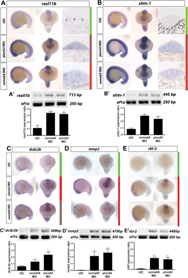
Microarray validation. In situ hybridization and RT-PCR validation of differential gene expression in plxnA2 and sema6A morphants at 18 somites. A–E: Top, middle, and bottom panels show uninjected control, plxnA2 MO injected, and sema6A MO injected embryos, respectively. Left and right panels show lateral and dorsal views, respectively, for each probe under each condition. Colored bars show corresponding heat map expression changes for each gene. UIC, uninjected control. A,B: Additional right panels show transverse 20 μm sections through 18 somite stage brain and eye vesicles processed for in situ hybridization with rasl11b and shtn-1 probes. Lines in whole-mounts indicate corresponding section locations. A′–E′: RT-PCR gels and quantification of gene expression changes for each gene as a ratio to ef1α control levels (rasl11b control 0.1137 ± 0.04, N = 3, rasl11b sema6A MO 0.34 ± 0.02, N = 3, **P < 0.01; rasl11b plxnA2 MO 0.32 ± 0.01, N = 3, **P < 0.01; shtn-1 control 0.08 ± 0.01, N = 3, shtn-1 sema6A MO 0.28 ± 0.01, N = 3, **P < 0.01; shtn-1 plxnA2 MO 0.24 ± 0.03, N = 3, **P < 0.01; dcdc2b control 0.06 ± 0.004, N = 3, dcdc2b sema6A MO 0.13 ± 0.02 N = 3, *P < 0.05; dcdc2b plxnA2 MO 0.24 ± 0.02, N = 3, ***P < 0.001; mmp2 control 0.004 ± 0.001, N = 5, mmp2 sema6A MO 0.21 ± 0.07, N = 5, *P < 0.05; mmp2 plxnA2 MO 0.27 ± 0.06, N = 5, **P < 0.01; rbl2 control 0.01 ± 0.002, N = 4, rbl2 sema6A MO 0.17 ± 0.02, N = 4, ***P < 0.001; rbl2 plxnA2 MO 0.15 ± 0.03, N = 4, **P < 0.01; one-way ANOVA, multiple comparisons test. Error bars indicate SEM). B, brain. E, eye. EXPRESSION / LABELING:
PHENOTYPE:
|
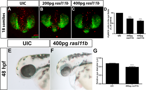
Overexpression of rasl11b decreases proliferation of RPCs. A–C: Confocal dorsal views of 18 somite rx3:GFP embryos co-labeled with pHH3. D: Increasing doses of rasl11b resulted in decreased proliferation within the GFP positive eye field (control 100%, N = 3, n = 30; 200 pg rasl11b 72.87% ± 4.25% N = 3, n = 38, ****P < 0.0001; 400 pg rasl11b 62.19% ± 4.01%, N = 3, n = 31, ****P < 0.0001; one-way ANOVA, multiple comparisons test; error bars indicate SEM). E–G: Overexpression of rasl11b results in smaller eyes at 48 hpf (eye diameter relative to head length; control 0.48 μm ± 0.005 μm N = 3, n = 30; 400 pg rasl11b 0.39 μm ± 0.01 μm, N = 3, n = 36; ****P < 0.0001; one-way ANOVA, multiple comparisons test; error bars indicate SEM). |