
- Title
-
The lim domain only protein 7 is important in zebrafish heart development
- Authors
- Ott, E.B., van den Akker, N.M., Sakalis, P.A., Gittenberger-de Groot, A.C., Te Velthuis, A.J., and Bagowski, C.P.
- Source
- Full text @ Dev. Dyn.

Gene expression patterns of LMO7 during zebrafish development. Expression results of whole-mount in situ hybridization are shown for the seven stages indicated. A-D,G,H,J: In additional to embryos between cleavage and end of gastrulation (A-D), we present dorsal views of embryos at 15som, prim-6, and long-pec stage (G,H,J). E,F, I: A higher magnification of the dorsal view at prim-6 (F) and a lateral (I) and frontal (E) view of the head at long-pec stage are shown. K,M: Close-ups of the heart at prim-6 and long-pec stage are presented. L,N: Additionally, sections of the hearts at these stages are shown. ba, branchial arches; di, diencephalon; fp, floorplate; h, heart; hb, hindbrain; mb, midbrain; mhb, midbrain-hindbrain boundary; ls, lens; ov, otic vesicle; pf, pectoral fin buds; pcl, proliferative cell layer; tct, tectum; tegm, tegmentum. |

Gene expression patterns of LMO7 during vertebrate development. Comparison of whole-mount in situ hybridization results of zebrafish, chicken and mouse embryos. Shown are three embryonic stages of each organism in lateral view. A,D,G: Zebrafish embryos are presented at prim-6, long-pec stage, and at 4 days postfertilization (dpf). B,E,H: For chicken, Hamburger-Hamilton stages HH18, HH20, and HH27 are shown. C,F,I: Mouse embryos are presented at 9.5, 10.5, and 11.5 days after gestation. ba, branchial arches; ce, cerebellum; di, diencephalon; fl, forelimb; h, heart; hl, hindlimb; int, intestine; ls, lens; lv, liver; la, liver anlage; lb, lung bud; ma, mandibular arches; mb, midbrain; mes, mesencephalon; met, metencephalon; mhb, midbrain-hindbrain boundary; myel, myelencephalon; ov, otic vesicle; pa, pharyngula arches; pf, pectoral fin buds; som, somites; tct, tectum; tel, telencephalon; tgm, tegmentum. |
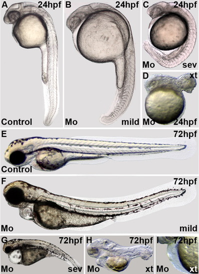
LMO7 morpholino injections. Effects of microinjection of LMO7 gene-specific morpholinos on zebrafish development. Shown are stereoscope pictures and DIC microscopy pictures of embryos and larvae at different stages of development. Embryos were injected at the one-cell stage with either a control morpholino or the LMO7 specific morpholinos (examples shown were injected with 4 ng/embryo of control-Mo or Mo-1). The magnifications are not in each case comparable between pictures. PHENOTYPE:
|
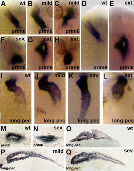
Effects of LMO7-morpholino injections on zebrafish heart development. Expression results of whole mount in situ hybridization of Cmcl2 in zebrafish embryos. Shown are zebrafish hearts of control injected and LMO7-morpholino injected embryos at prim-6 and long-pec stages. For morpholino-injected embryos mild, severe and extreme phenotypes are presented. A, B, C, F, G, H: Effects of the microinjection on zebrafish hearts at prim-6 are shown. D and E: Additionally, we present effects on heart localization. M, N: Also sections of zebrafish hearts at prim-6 are presented. I-L: Hearts of control and LMO7 injected embryos are shown. For the latter, the hearts of embryos with mild, severe and extreme phenotypes are depicted. Furthermore, longitudinal sections of hearts at the long pec stage are presented for wildtype (wt) embryos and morpholino-injected embryos showing a mild and severe phenotype (O-Q). |
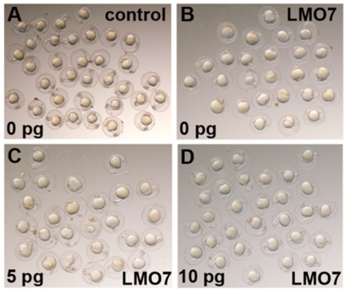
Examples of control Morpholino (A) and LMO7 Mo (B-D) injected embryos. Embryos were injected at the 1-2 cell stage and are shown at 24 hpf. In (C) and (D) LMO7 RNA was coinjected (5 pg and 10 pg, respectively). Examples show 35 embryos in total in (A) with no morphants. In (B) 25 embryos including 14 morphants, in (C) 25 embryos including 4 morphants and in (D) 30 embryos including 4 morphants are depicted. Embryos were randomly selected and photographed by stereomicroscopy. The results of all rescue experiments are shown in table 4. PHENOTYPE:
|
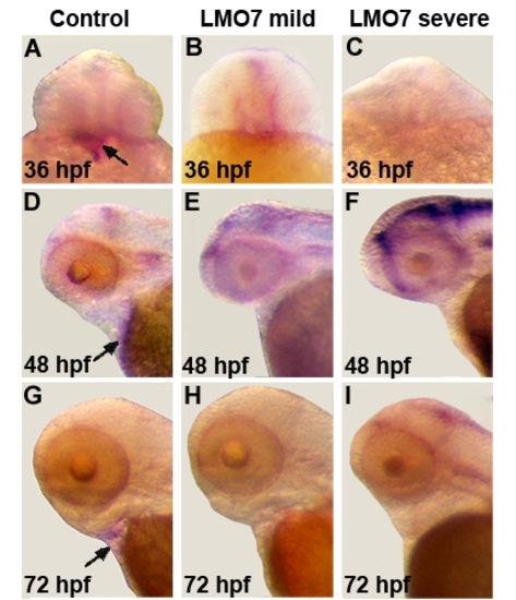
In situ hybridization using a notch1b probe (A-I). Control embryos show staining in the heart (arrows in A,D and G), whereas LMO7 knock down embryos don't. Examples shown in B,E and H (mild) and C,F and I (severe) Heart staining was never observed in LMO7 knock down embryos. EXPRESSION / LABELING:
PHENOTYPE:
|

Unillustrated author statements PHENOTYPE:
|