
- Title
-
Localization of anosmin-1a and anosmin-1b in the inner ear and neuromasts of zebrafish
- Authors
- Ernest, S., Guadagnini, S., Prevost, M.C., and Soussi-Yanicostas, N.
- Source
- Full text @ Gene Expr. Patterns

Organization of the adult zebrafish inner ear. (A) Scheme of an adult zebrafish inner ear showing all sensory epithelia (in red). The three main macula are the utricule (u), the saccule (s) and the lagena (l), the fourth, smaller one, is the macula neglecta (mn); the three crista of the semi-circular canals are the anterior cristae (ac), the lateral cristae (lc), and the posterior cristae (pc). (B–C) Micro-dissected adult zebrafish inner ear. Whole adult inner ear showing the localization of the three main macula and of the three crista, as well as innervation of the anterior cristae (B) (note that the semi-circular canals were partially broken during hand dissection). Detail of the anterior portion of the inner ear, showing the anterior and the lateral crista together with the innervation of the lateral cristae (C). (D) Schematic representation of a cristae ampullaris with a cross section at one extremity of the cristae; the colors used in this scheme correspond to the colors shown in immunofluorescence experiments (hair cell body, kinocilium and stereocilia are depicted in green, blue, and red, respectively ; ampullary nerves are shown in blue). (E–F) Sensory patches of the anterior part of the inner ear. The anterior and lateral crista and the utricular maculae depicted here were stained with rhodamine–phalloidin and microphotographed using Nomarski optics (E) or a red fluorescence filter (F). The lateral cristae is shown longitudinally and the anterior cristae transversally. |
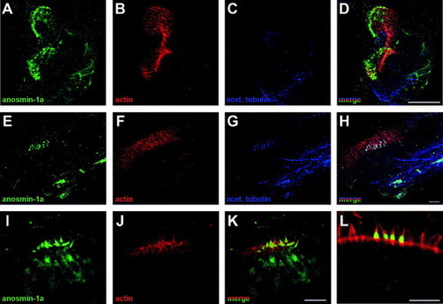
Expression of anosmin-1a in sensory hair cells of the cristae and in the ampullary nerve of adult zebrafish inner ear. Whole-mount double immunofluorescence with anti-anosmin-1a polyclonal (green) and anti-acetylated tubulin monoclonal (blue) antibodies combined to rhodamin-phalloidin staining of actin (red). (A–D) In the whole cristae, anosmin-1a staining was observed at both extremities of the sensory patch and in the ampullary nerve. (E–H) At the extremity of the cristae, anosmin-1a is accumulated in the ciliated cells, mainly in the hair bundle but also in the cell body, and in the nerve fibers. (I–K) Higher magnification revealed anosmin-1a accumulation in the actin-rich hair bundle and also in the cell body. (L) Confocal section showing anosmin-1a staining of the cilia, in the supranuclear region and at the cell membrane. Data are maximum intensity projections of the acquired z-stacks (A–K) or a single confocal section (L). Scale bar: 50 μm (A–D), 10 μm (E–L). EXPRESSION / LABELING:
|

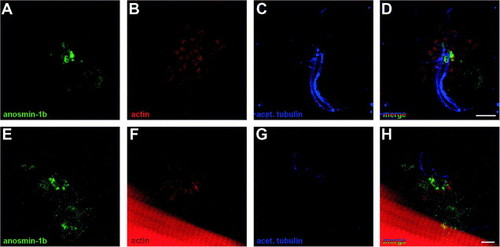
Expression of anosmin-1b in sensory hair cells of the cristae of adult zebrafish inner ear. (A–D) Imaging of the whole cristae (longitudinal view) showing anosmin-1b staining (green) at both extremities of the sensory patch. No anosmin-1b expression (anti-anosmin-1b polyclonal antibody) was detected in the nerve, labeled with a monoclonal anti-acetylated tubulin antibody (blue). (E–G) Pattern of accumulation of anosmin-1b at one extremity of the cristae (transversal view). (H–J) Detail of one extremity of the cristae showing anosmin-1b accumulation in the body and hair bundle of the sensory cells. Data are maximum intensity projections of the acquired z-stacks (A–J). Scale bar: 50 μm (A–D), 10 μm (E–J). EXPRESSION / LABELING:
|

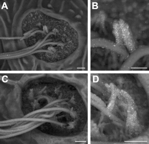
The lateral line of a 120 hpf zebrafish embryo. (A–D) Scanning electron microscopy (SEM) of the lateral line neuromasts. Neuromasts are indicated by arrowheads (A). Magnified view of the area squared in (A) showing the three terminal neuromasts localized at the tip of the tail (B). View of a neuromast, with the kinocilia and stereocilia of the sensory cells pointing out of the organ. (C). Higher magnification of the neuromast pictured in (C) showing the hair bundles (stereocilia) and the anchoring point of the kinocilium of three hair cells (D). Note that the long kinocilia have been pushed against the body of the fish during sample preparation. Scale bar: 100 μm (A), 50 μm (B), 1 μm (C–D). (E) Schematic representation of the sensory hair cell of the neuromast. Hair cells are depicted within the ultrastructure of the neuromast on the left side. On the right side is shown a detail of the apical surface of a sensory hair cell, with the kinocilium and the stereocilia arranged in staircase. (Ghysen, A., Dambly–Chaudiere, C., 2004. Simplified scheme adapted from development of the zebrafish lateral line. Current Opinion in Neurobiology 14, 67–73). The colors used in the scheme correspond to the colors observed in the immunofluorescence experiments shown in Fig. 5 and Fig. 6. |
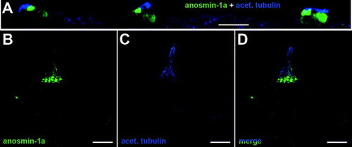
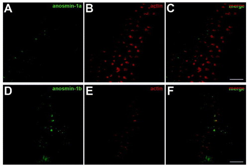
Expression of anosmin-1a in sensory hair cells of the neuromast. (A) Confocal sections of the three terminal neuromasts showing anosmin-1a staining (green) in the hair bundle and, at a lower level, in the body of sensory hair cells. Kinocilia were labeled with an anti-acetylated tubulin antibody (blue). (B–D) Higher magnification of a single neuromast revealed a strong anosmin-1a accumulation in the stereocilia of hair cells and, to a lesser extent, in the cell body and in the kinocilia (lower half) which was co-labeled using an anti-acetylated tubulin antibody. Data represent the maximum intensity projection of the z-sections of the acquired z-stacks (A) or maximum intensity projections of the acquired z-stacks (B–D). Scale bar: 20 μm (A), 10 μm (B–D). |
Expression of anosmin-1b in neuromast sensory hair cells. 120 hpf embryos were subjected to double immunofluorescence with anti-anosmin-1b polyclonal (green) and anti-acetylated tubulin monoclonal (blue) antibodies combined to TRITC-conjugated phalloidin staining of actin (red). (A–D) Confocal imaging of a lateral line neuromast, performed according to the apico-basal axis of hair cells, revealed anosmin-1b accumulation in the central most sensory hair cells. Projection of the confocal sections demonstrated staining in the hair bundle and, more weakly, at the cell membrane. (E–H) Projection of transverse confocal sections of a neuromast showing the apico-basal distribution of anosmin-1b within hair cells, with a dense staining observed at the top portion of the stereocilia and a weaker signal at the cell membrane. Data are maximum intensity projections of the acquired z-stacks. Scale bar: 5 μm (A–H). |
Scanning electron microscopic detection of anosmin-1a and anosmin-1b immunogold–labeling in neuromasts of 120 hpf zebrafish embryos. (A–B) A high level of anosmin-1a staining was detected on the stereocilia and, to a lesser extent, on the kinocilia of neuromasts hair cells. (C–D) anosmin-1b accumulation was also observed on the stereocilia and, at a lower level, on the kinocilia of neuromasts hair cells. Scale bar: 1 μm (A, C), 500 nm (B, D). |
|
Reprinted from Gene expression patterns : GEP, 7(3), Ernest, S., Guadagnini, S., Prevost, M.C., and Soussi-Yanicostas, N., Localization of anosmin-1a and anosmin-1b in the inner ear and neuromasts of zebrafish, 274-281, Copyright (2007) with permission from Elsevier. Full text @ Gene Expr. Patterns