
Fig. 3
- ID
- ZDB-IMAGE-071001-111
- Genes
- Publication
- Ernest et al., 2007 - Localization of anosmin-1a and anosmin-1b in the inner ear and neuromasts of zebrafish
- All Figures
- Figures for Ernest et al., 2007
|
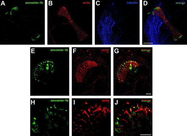
Fig. 3 Expression of anosmin-1b in sensory hair cells of the cristae of adult zebrafish inner ear. (A–D) Imaging of the whole cristae (longitudinal view) showing anosmin-1b staining (green) at both extremities of the sensory patch. No anosmin-1b expression (anti-anosmin-1b polyclonal antibody) was detected in the nerve, labeled with a monoclonal anti-acetylated tubulin antibody (blue). (E–G) Pattern of accumulation of anosmin-1b at one extremity of the cristae (transversal view). (H–J) Detail of one extremity of the cristae showing anosmin-1b accumulation in the body and hair bundle of the sensory cells. Data are maximum intensity projections of the acquired z-stacks (A–J). Scale bar: 50 μm (A–D), 10 μm (E–J).
Reprinted from Gene expression patterns : GEP, 7(3), Ernest, S., Guadagnini, S., Prevost, M.C., and Soussi-Yanicostas, N., Localization of anosmin-1a and anosmin-1b in the inner ear and neuromasts of zebrafish, 274-281, Copyright (2007) with permission from Elsevier. Full text @ Gene Expr. Patterns