
FIGURE
Fig. 7
- ID
- ZDB-FIG-071001-115
- Publication
- Ernest et al., 2007 - Localization of anosmin-1a and anosmin-1b in the inner ear and neuromasts of zebrafish
- Other Figures
- All Figure Page
- Back to All Figure Page
Fig. 7
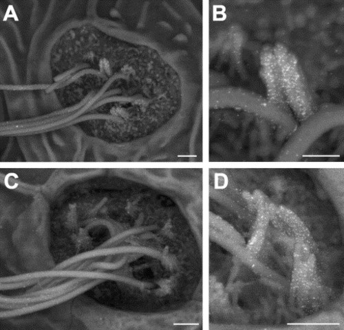
Scanning electron microscopic detection of anosmin-1a and anosmin-1b immunogold–labeling in neuromasts of 120 hpf zebrafish embryos. (A–B) A high level of anosmin-1a staining was detected on the stereocilia and, to a lesser extent, on the kinocilia of neuromasts hair cells. (C–D) anosmin-1b accumulation was also observed on the stereocilia and, at a lower level, on the kinocilia of neuromasts hair cells. Scale bar: 1 μm (A, C), 500 nm (B, D). |
Expression Data
Expression Detail
Antibody Labeling
Phenotype Data
Phenotype Detail
Acknowledgments
This image is the copyrighted work of the attributed author or publisher, and
ZFIN has permission only to display this image to its users.
Additional permissions should be obtained from the applicable author or publisher of the image.
Reprinted from Gene expression patterns : GEP, 7(3), Ernest, S., Guadagnini, S., Prevost, M.C., and Soussi-Yanicostas, N., Localization of anosmin-1a and anosmin-1b in the inner ear and neuromasts of zebrafish, 274-281, Copyright (2007) with permission from Elsevier. Full text @ Gene Expr. Patterns