
Fig. 3
|
Fig. 3
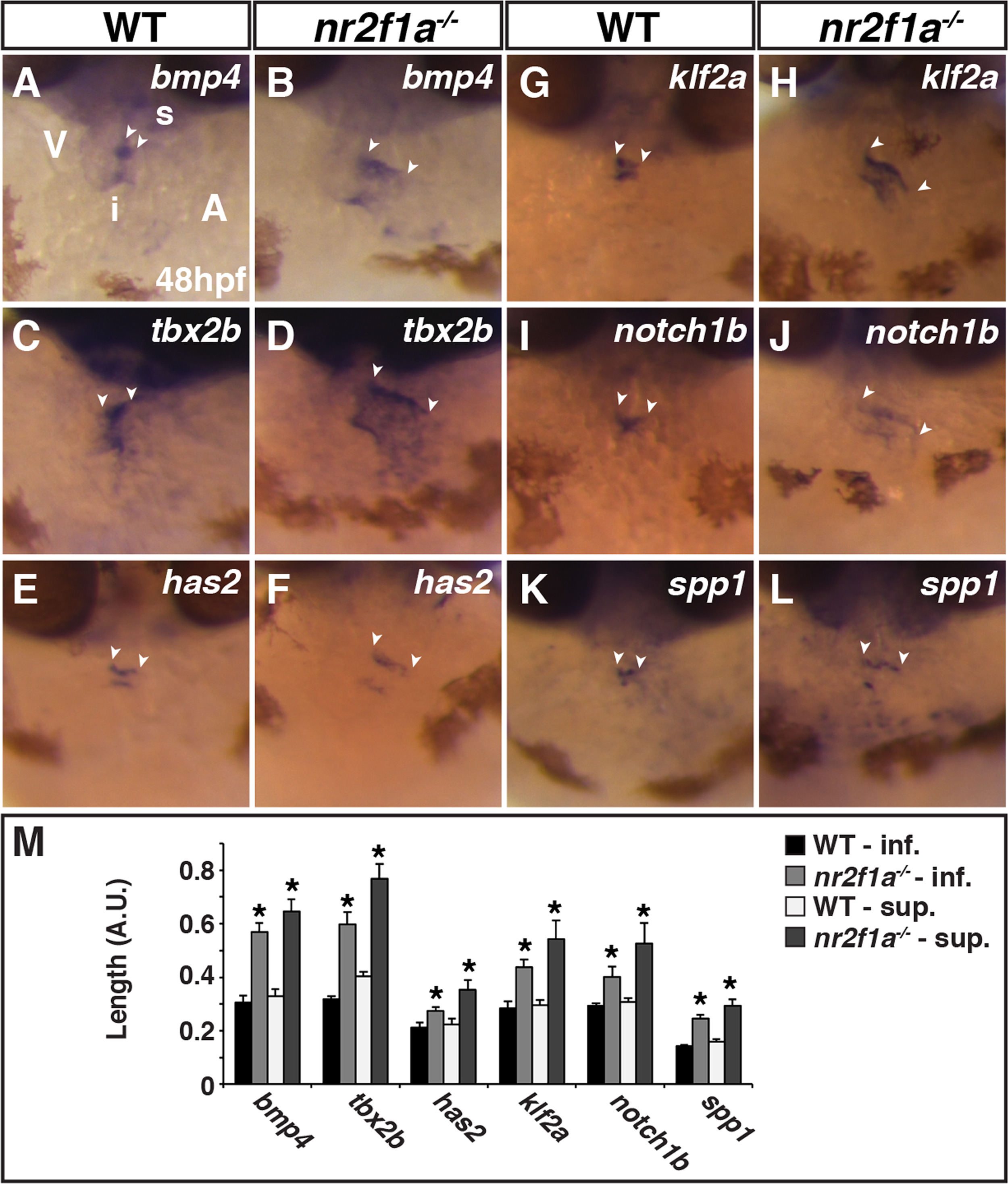
AVC markers are expanded innr2f1amutant hearts. (A-L) ISH for bmp4 (A,B), tbx2b (C,D), has2 (E,F), klf2a (G,H), notch1b (I,J), and spp1 (K,L) in WT and nr2f1a mutant embryos. Images are frontal views with dorsal up. The relative position of the ventricle and atrium is the same in all images. Arrowheads indicate the length of expression on the superior side (s) of the AVC, which is adjacent to the inner curvature of the ventricle. Inferior side (i) of the AVC is adjacent to the outer curvature of the ventricle. (M) Length measurements of marker expression in the AVC. (WT bmp4 n = 7, nr2f1a-/-bmp4 n = 7, WT tbx2b n = 10, nr2f1a-/-tbx2b n = 10, WT has n = 7, nr2f1a-/-has n = 7, WT klf2a n = 6, nr2f1a-/-klf2a n = 6, WT notch1b n = 5, nr2f1a-/-notch1b n = 5, WT spp1 n = 9, nr2f1a-/-klf2a n = 9.).
Reprinted from Developmental Biology, 434(1), Duong, T.B., Ravisankar, P., Song, Y.C., Gafranek, J.T., Rydeen, A.B., Dohn, T.E., Barske, L.A., Crump, J.G., Waxman, J.S., Nr2f1a balances atrial chamber and atrioventricular canal size via BMP signaling-independent and -dependent mechanisms, 7-14, Copyright (2017) with permission from Elsevier. Full text @ Dev. Biol.