
Fig. 2
|
Fig. 2
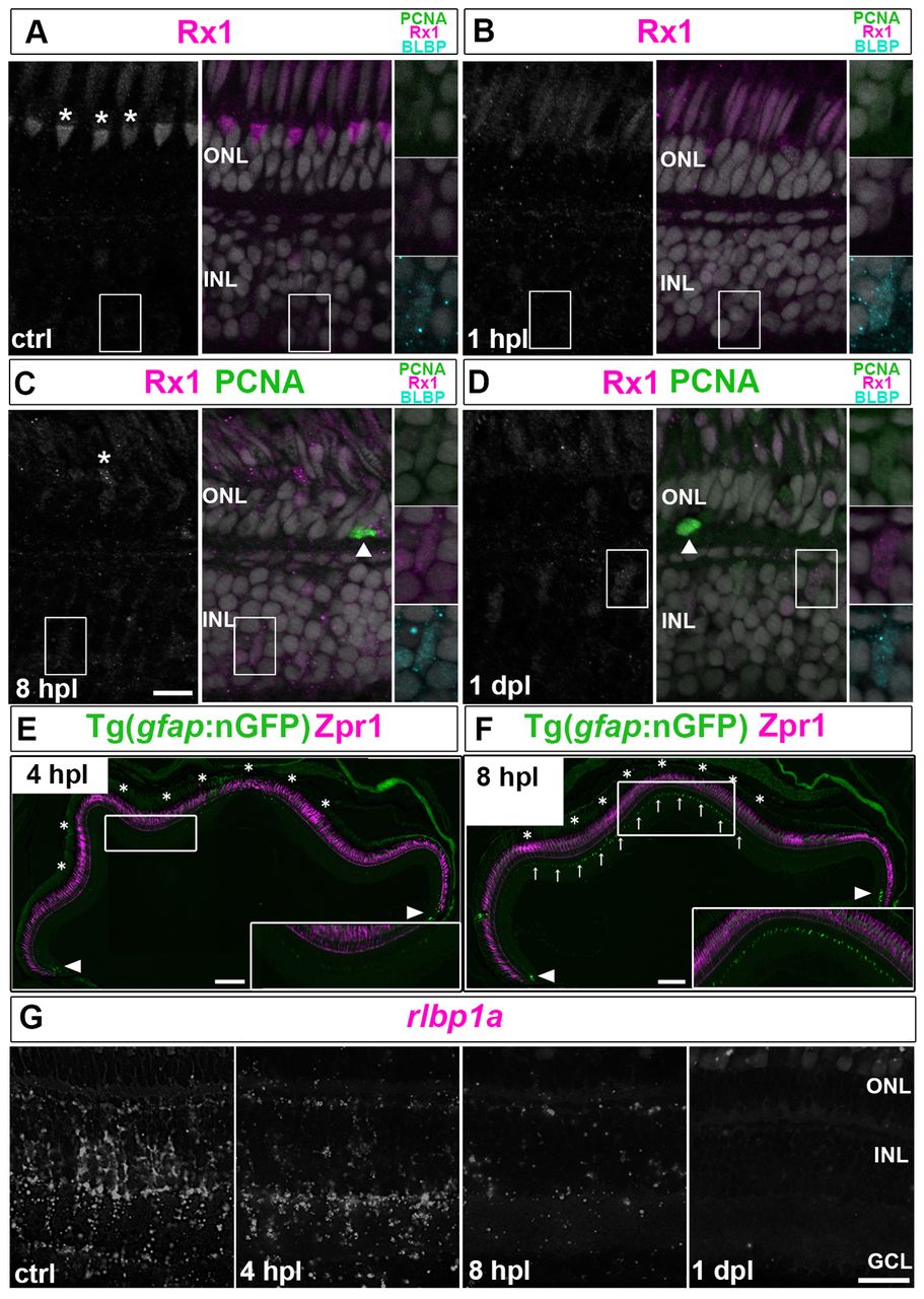
Müller glia partially dedifferentiate and express retinal progenitor markers in response to photoreceptor lesions. (A-D) PCNA (green), BLBP (cyan) and Rx1 (white/magenta) in control (ctrl) retina. Higher magnifications of the boxed regions are shown on the right. (A) Cones are Rx1+ (asterisks, UV cone nuclei). (B) By 1 hpl, Rx1 is reduced in UV cones and BLBP is upregulated in Müller glia. (C) At 8 hpl, cones collapse (asterisk) and Müller glia express BLBP and Rx1 (boxed region). A PCNA+ microglia or rod precursor (arrowhead) can be seen in the ONL. (D) At 1 dpl, Müller glia are weakly PCNA+/BLBP+/Rx1+. (E,F) Injury-induced nGFP in lesioned area (asterisks) at 4 and 8 hpl and in immature Müller glia (arrowheads). Zpr1 (magenta), which recognizes Arrestin 3a and specifically labels red/green cones. Higher magnifications of the boxed regions are shown in insets. (C) Reduced rlbp1a transcript (white) by 4 hpl and no detectable signal at 1 dpl. Scale bars: 10 μm (A-D,G); 100 μm (E,F).