
|
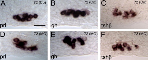
Fig. 5
ISH on transverse sections of controls (Co; A–C) and prop1 MO (MO; D–F) injected embryos at 72 hpf using RNA probes for the pituitary lineage specific markers prl gh and tshβ. All three markers were strongly expressed in the pituitaries of both control and morphant embryos. Impaired cellular organization was observed for lactotrope (D vs. A) and somatotrope (E vs. B) lineages while thyrotropes were apparently unaffected by prop1 deficiency (F vs. C). Age in hpf and (treatments) are indicated in the upper-right hand corner; gene names in the lower-left corner. All panels are equally scaled (scale bar = 25 μm).