
IMAGE
Fig. 5
Image
|
Figure Caption
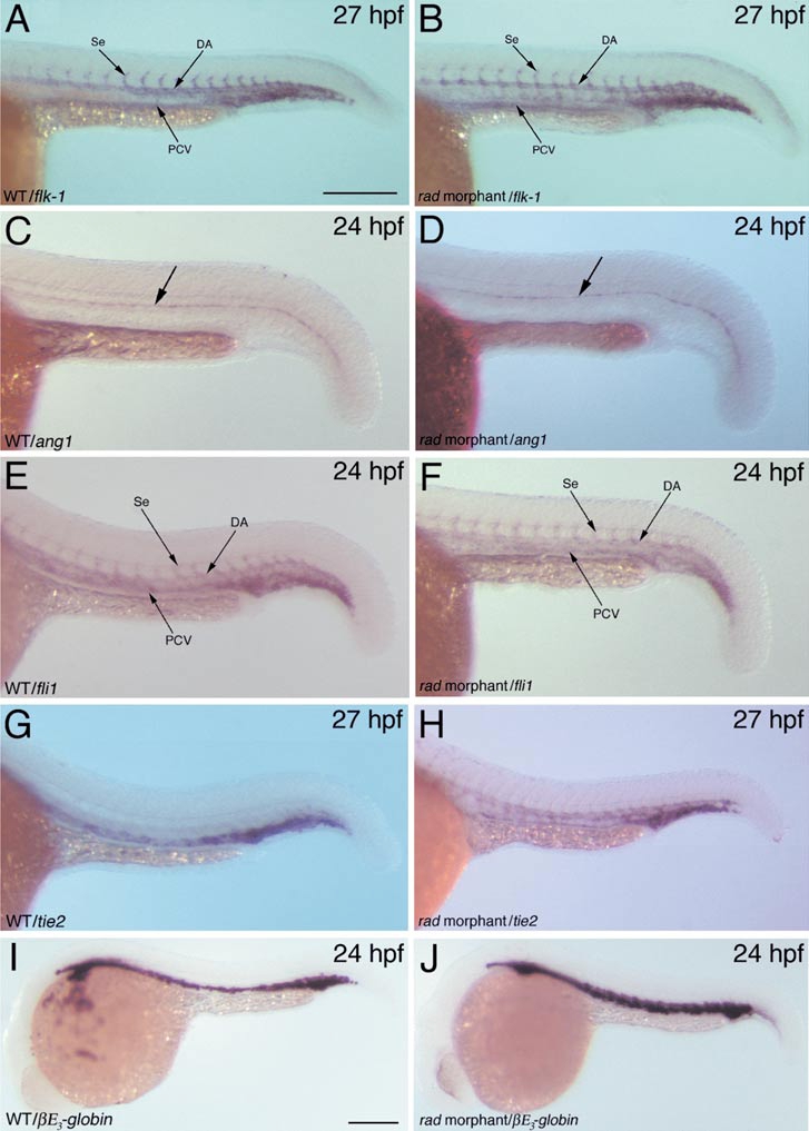
Fig. 5 Normal vascular patterning and hematopoiesis in radar morphant embryos. Whole-mount in situ hybridization analysis of flk-1 (A, B), ang1 (C, D), fli1 (E, F), tie2 (G, H), and βE3-globin (I, J) in wild type (A, C, E, G, I) and radar morphant (B, D, F, H, J; injected with 8 ng rad-MO1) embryos. DA, dorsal aorta; PCV, posterior cardinal vein; Se, intersegmental vessel. Scale bars in (A) and (I) represent 250 and 200 μm, respectively.
Figure Data
Acknowledgments
This image is the copyrighted work of the attributed author or publisher, and
ZFIN has permission only to display this image to its users.
Additional permissions should be obtained from the applicable author or publisher of the image.
Reprinted from Developmental Biology, 251(1), Hall, C.J., Flores, M.V.C., Davidson, A.J., Crosier, K.E., and Crosier, P.S., Radar is required for the establishment of vascular integrity in the zebrafish, 105-117, Copyright (2002) with permission from Elsevier. Full text @ Dev. Biol.