
|
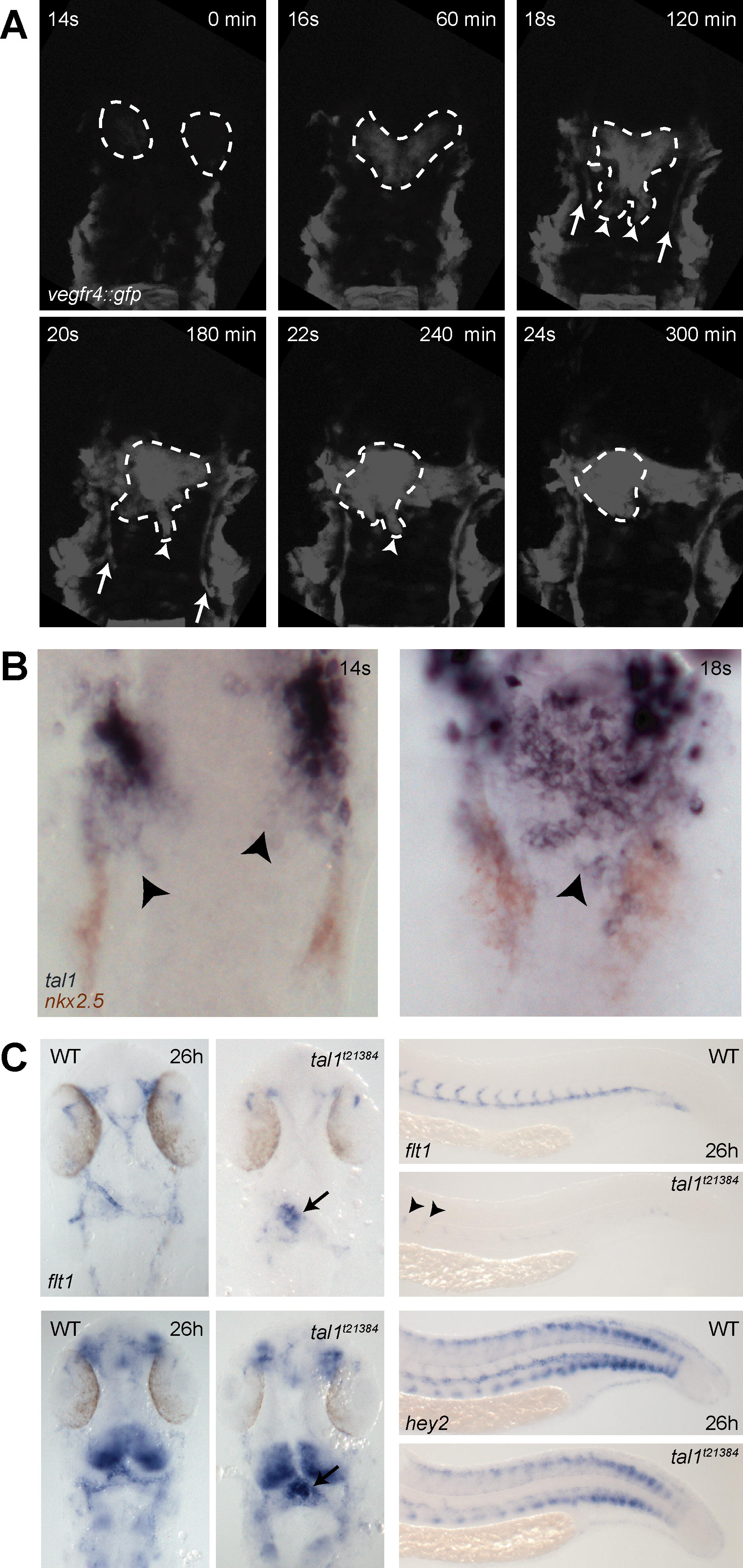
Fig. 5 Migration of Endocardial Precursors and Heart Tube Formation in wt tal1t21384 Mutant Embryos (A) tal121384 mutant embryos transgenic for vegfr4:gfp were subjected to time-lapse confocal microscopy, revealing defects during early endocardial precursor migration. A movie demonstrating this process can be viewed in the Video S3. Six individual frames from this movie are shown in (A). Whereas the initial formation of bilateral endocardial precursors is not affected, posterior migration is disturbed, and endocardial precursors remain attached in a relative anterior position. Note that migration of the paired lateral dorsal aortae (arrows) proceeds normally. (B) tal1 expression in endocardial precursors. Two-color in situ hybridization revealing tal1 (blue) and nkx2.5 (red) expression in wt embryos at the 14- and 18-somite stage. tal1 expression is observed in endocardial but not myocardial precursors during their posterior migration (arrowheads). (C) Expression of the arterial markers flt1 and hey2 is retained in tal1t21384 mutant endocardium, but severely reduced in endothelium, as revealed by in situ hybridization. Dorsal view of flt1 and hey2 expression in the head, anterior to the top, and lateral view of flt1 and hey2 in the tail (28 hpf). In wt embryos, flt1 expression is observed in all head arteries, the aortic arches and the endocardium (arrow). In tal1t21384 mutant embryos, expression of flt1 is observed in a few remaining head arteries and the aortic arches. High levels are seen in the endocardium (arrow). In wt embryos, flt1 expression is observed in the dorsal aorta and the developing intersegmental vessels. In the tail of tal1t21384 mutant embryos, expression of flt1 is abolished, except for a few remaining cells that express flt1 at low levels (arrowheads). In wt embryos, hey2 expression is observed in the endocardium and the aortic arches and in some parts of the brain and spinal chord. In tal1t21384 mutant embryos, expression in the endocardium (arrow) is increased. In wt embryos, hey2 expression is observed in the dorsal aorta and the developing intersegmental vessels, spinal chord neurons, and in ventral and dorsal cells of the somites. In tal1t21384mutant embryos, expression in the dorsal aorta and intersegmental vessels is severely reduced, although some anterior intersegmental vessels and aortic cells retain low levels of hey2 expression (arrowheads).