
|
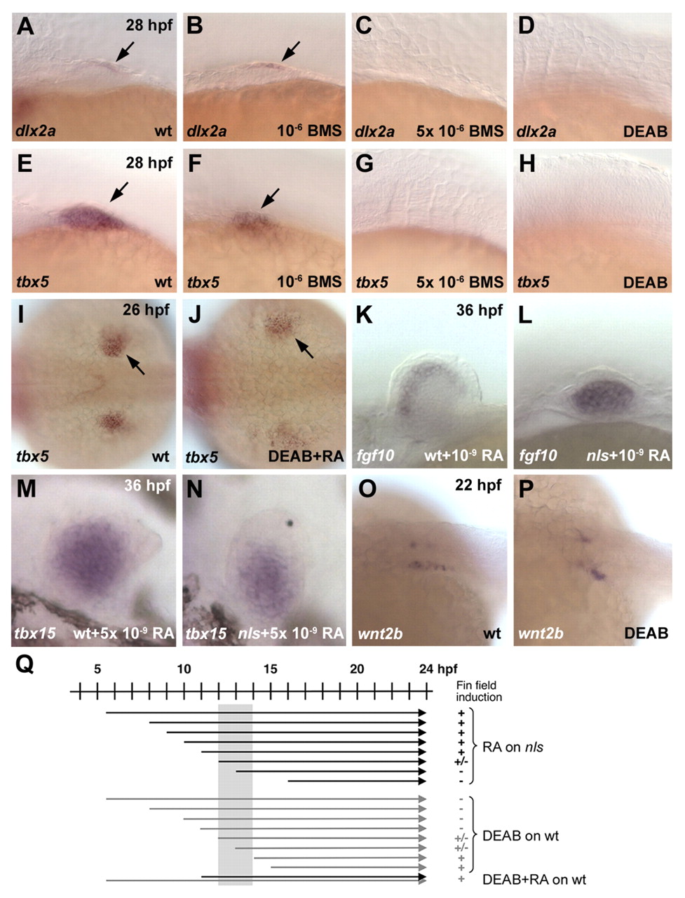
Fig. 1 Pectoral fin field induction requires retinoic acid signaling during somitogenesis. Anterior is towards the left: lateral (A-H,K-N), dorsal (I,J) and dorsolateral (O,P) views. Whole mount in situ hybridizations. (A-H) Wild-type embryos treated from 10 hpf onwards with 10-6 M and 5x10⁻⁶ M BMS493 (B,F and C,G) and with 10 µM DEAB(D,H); untreated controls (A,E). Expression of mesenchymal (tbx5) and apical epidermal fold (dlx2a) markers (arrows) is lost at the higher antagonist concentration and in DEAB treatments. (I,J) In the absence of RA synthesis from 5 hpf onwards (10 µM DEAB), tbx5 expression is restored to near-normal levels by treatment with 10-8 M RA from 11 hours onwards (arrows) (J). (K-N) 10-9 M RA (11-36 hpf) rescues fgf10 expression (K,L) and 5x10⁻⁹ M RA (11-36 hpf) rescues tbx15 expression (M,N) in nls. (O,P) wnt2b expression is not abolished in wild-type animals treated with DEAB from 11 hpf onwards. (Q) Schematic overview of the treatment applied to wild-type embryos depicted in A-J; shaded area depicts time window in which RA signaling is required.