
FIGURE
Fig. 1
- ID
- ZDB-FIG-240620-118
- Publication
- Hiraki-Kajiyama et al., 2024 - An atlas and database of neuropeptide gene expression in the adult zebrafish forebrain
- Other Figures
- All Figure Page
- Back to All Figure Page
Fig. 1
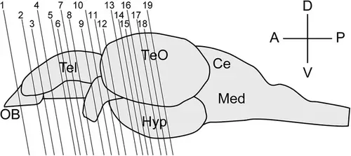
Schematic of the zebrafish brain viewed from the lateral side. The anterior is to the left, and the dorsal is to the top. Lines 1−19 indicate the locations of each plane in Figures 2-20. OB, olfactory bulb; TeO, optic tectum; Hyp, hypothalamus; Ce, cerebellum; Med, medulla oblongata; A, anterior; P, posterior; D, dorsal; V, ventral. |
Expression Data
Expression Detail
Antibody Labeling
Phenotype Data
Phenotype Detail
Acknowledgments
This image is the copyrighted work of the attributed author or publisher, and
ZFIN has permission only to display this image to its users.
Additional permissions should be obtained from the applicable author or publisher of the image.
Full text @ J. Comp. Neurol.