
Fig. 4
- ID
- ZDB-FIG-231215-118
- Publication
- Schiavo et al., 2021 - Vascular endothelial growth factor-c regulates hematopoietic stem cell fate in the dorsal aorta
- Other Figures
- All Figure Page
- Back to All Figure Page
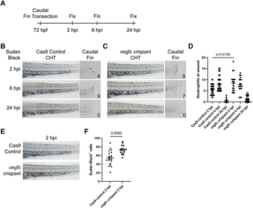
vegfc loss-of-function embryos show altered injury response. (A) Experimental approach. Caudal fins were transected distally without injury of the notochord at 72 hpf. Embryos were fixed in 4% PFA at different intervals following transection. (B) Bright-view images of Sudan Black staining in Cas9 control embryos depicting neutrophil response at 2, 6 and 24 hpi. Neutrophil response peaks at 6 hpi and subsides by 24 hpi. (C) Bright-view images of Sudan Black staining in vegfc loss-of-function embryos. Neutrophil response peaks at 2 hpi and diminishes over time. (D) Quantification of neutrophil number within the injury area, P=0.0138 (comparison between Cas9 control 2 hpi and vegfc crispant 2 hpi; one-way ANOVA). (E) Representative images showing increased neutrophils within the CHT of vegfc loss-of-function embryos at 2 hpi. (F) Quantification of the number of Sudan Black-positive neutrophils within the CHT at 2 hpi, P=0.0003 (unpaired two-tailed t-test). Error bars show mean±s.e.m. |