
Fig. 1
- ID
- ZDB-FIG-231215-115
- Publication
- Schiavo et al., 2021 - Vascular endothelial growth factor-c regulates hematopoietic stem cell fate in the dorsal aorta
- Other Figures
- All Figure Page
- Back to All Figure Page
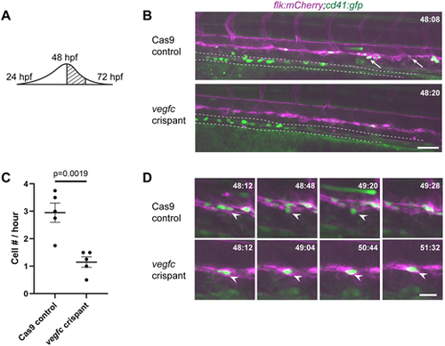
vegfc loss-of-function results in decreased HSPC emergence from the DA. (A) Schematic showing the wave of emergence from the DA; this experiment occurs during the peak. (B) Single frame of time-lapse (hpf:min) showing cd41:gfp+ (green) HSPCs (arrows) budding from flk:mCherry+ (magenta) HE in Cas9 control (top panel) or vegfc crispant embryos (bottom panel). Dotted line outlines developing pronephric tubules (see Movies 1 and 2). (C) Quantification of the total number of cd41:gfp+ HSPCs budding from the DA between 48 and 52 hpf, divided by four to give cell number (#) per hour. n=5 embryos per condition, P=0.0019 (unpaired two-tailed t-test). (D) Four frames selected from Cas9 control and vegfc crispant time-lapse movies depicting HSPC budding (top panels) and a static hammock-like cell (bottom panels). White arrowheads indicate the cell of interest. Error bars show mean±s.e.m. Scale bars: 50 µm (B); 10 µm (D). |