
FIGURE
Figure 2
- ID
- ZDB-FIG-221226-315
- Publication
- Bandla et al., 2022 - A New Transgenic Tool to Study the Ret Signaling Pathway in the Enteric Nervous System
- Other Figures
- All Figure Page
- Back to All Figure Page
Figure 2
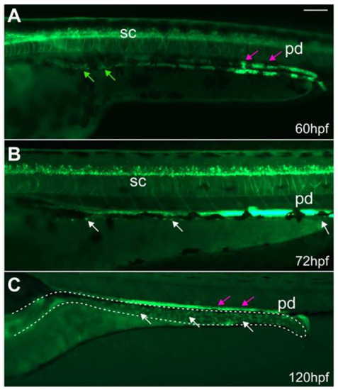
ret:GFP+ EPCs migrate along the developing gut and populate the gut. At (A) 60 hpf, GFP+ EPCs (green arrows) migrate along the developing gut. At (B) 72 and (C) 120 hpf, GFP+ ENS cells (white arrows) populate the gut (dashed line). Some of the GFP+ cells in the gut are enteroendocrine cells. The GFP+ pronephric ducts (pd, magenta arrows) directly overlay the ENS. (A–C): Whole-mount side-views of embryos/larvae at the stage indicated. Dashed line outlines the gut. Scale bar = 100 µm. |
Expression Data
| Gene: | |
|---|---|
| Fish: | |
| Anatomical Terms: | |
| Stage Range: | Protruding-mouth to Day 5 |
Expression Detail
Antibody Labeling
Phenotype Data
Phenotype Detail
Acknowledgments
This image is the copyrighted work of the attributed author or publisher, and
ZFIN has permission only to display this image to its users.
Additional permissions should be obtained from the applicable author or publisher of the image.
Full text @ Int. J. Mol. Sci.