
Figure 1
- ID
- ZDB-FIG-221226-314
- Publication
- Bandla et al., 2022 - A New Transgenic Tool to Study the Ret Signaling Pathway in the Enteric Nervous System
- Other Figures
- All Figure Page
- Back to All Figure Page
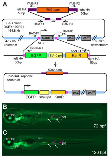
Overview of BAC-construct cloning strategy. (A) The start codon and exon 1 of ret were replaced by homologous recombination with the EGFP-SV40-pA-KanR construct using 50 bp-long homology arms (HA, magenta box) as indicated. iTol2 sites were included in the BAC backbone as shown using homologous recombination. The arrows indicate forward and reverse primer pairs to verify the correct generation of the tol2-BAC reporter construct. Integration of the BAC DNA does not result in overexpression of ret as the ret ATG is replaced by the EGFP cassette and the EGFP insert contains a strong transcription termination signal (SV40 pA, simian virus 40 poly A) [30]. Overview of GFP+ cells in the retina, pharyngeal arches (pa), spinal cord (sc), pronephric duct (pd, magenta arrows), and ENS cells (white arrows) at 72 (B) and 120 (C) hours post fertilization (hpf). (B,C): Whole-mount side-views of embryo/larva at the stage indicated. KanR kanamycin. Scale bar = 200 µm. |