
Fig. 2
- ID
- ZDB-FIG-221222-28
- Publication
- Terai et al., 2021 - Electrophysiological and pharmacological characterization of spreading depolarization in the adult zebrafish tectum
- Other Figures
- All Figure Page
- Back to All Figure Page
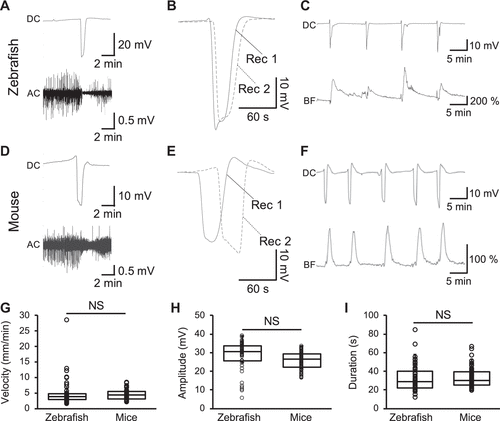
Figure 2.Concurrent changes in alternating current (AC) potential and hemodynamics of the zebrafish tectum during spreading depolarization (SD). Representative traces of the direct current (DC, top) and AC (bottom) components of the extracellular potential recorded in the zebrafish tectum (A) and the mouse cortex (D) after KCl stimulation. Representative traces of the DC potential recorded with two electrodes (Rec1 and Rec2) in the zebrafish tectum (B, interelectrode distance: 0.5 mm) and mouse cortex (E, interelectrode distance: 2 mm). Note the delayed negative peak in Rec2, which is further from the stimulation site than Rec1. Representative traces of the DC potential (top) and local blood flow (bottom) in the zebrafish tectum (C) and the mouse cortex (F) upon KCl stimulation. Bar graphs with dot-whisker plots of the velocity (G), amplitude (H), and duration at the half of the maximal amplitude (I) of SD recorded in the zebrafish tectum and the mouse cortex. NS, not significant; n = 8 fish, n = 4 mice. |