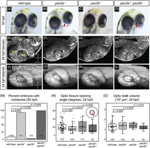
pax2a and pax2a; pax2b loss‐of‐function mutants display coloboma, but optic cup morphogenesis appears normal. (A‐D) Eye phenotypes at 55 hpf. (A) Wild‐type embryo. (B) pax2a tu29a mutant embryo; coloboma is apparent as a region of hypopigmentation at the back of the eye (red arrowhead). (C) pax2b sa10953 mutant embryo; the eye is evenly pigmented, there is no apparent coloboma. (D) pax2a tu29a ; pax2b sa10953 mutant embryo; coloboma is apparent (red arrowhead). (E‐L) Optic cup phenotypes at 24 hpf. (E) Wild‐type, (F) pax2a tu29a mutant, (G) pax2b sa10953 mutant, (H) and pax2a tu29a ; pax2b sa10953 mutant optic cup formation, single confocal slices. Dorsal view. Cell membranes, grayscale [Tg(bactin2:EGFP‐CAAX)]. (I) Wild‐type, (J) pax2a tu29a mutant, (K) pax2b sa10953 mutant, (L) and pax2a tu29a ; pax2b sa10953 mutant optic cup formation, three‐dimensional rendering. Lateral view. Cell membranes, grayscale [Tg(bactin2:EGFP‐CAAX)]. Yellow dashed lines indicate optic fissure margins. (M) Penetrance of coloboma phenotype, 55 hpf. n (embryos) shown at base of graphs. (N) Quantification of optic fissure opening angle measurement, 24 hpf. n (embryos) shown at base of graphs. Schematic depicts the optic fissure opening angle measurement in which each ray (red) originates at the margins of the optic fissure and the vertex lies in the center of the lens. (O) Quantification of optic stalk volume, 24 hpf. n (embryos) shown at base of graphs. P‐values for (M‐O) were calculated using an unpaired Student's t‐test. br, brain; le, lens; nr, neural retina. Scale bar: 50 μm
|
