
Fig. 1
- ID
- ZDB-FIG-220808-56
- Publication
- Lusk et al., 2021 - Pax2a, but not pax2b, influences cell survival and periocular mesenchyme localization to facilitate zebrafish optic fissure closure
- Other Figures
- All Figure Page
- Back to All Figure Page
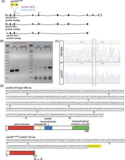
The pax2b sa10953 allele contains an essential splice site variant causing retention of intron 3/4 and a premature stop codon. (A) Using the zebrafish GRCz11 genome assembly, the pax2b gene is found on Chromosome 12: 45,799,982 to 45,876,387. Schematized are three predicted transcripts; the pax2b sa10953 allele contains a G > A mutation at site Chr 12:45872685 that is predicted to affect all transcripts. This schematic is shown reflected compared to the genomic organization for ease of interpretation, as pax2b lies on the reverse strand. Untranslated regions are depicted as white boxes, exons as red boxes, and introns as connecting lines. Two primer pairs are represented as arrows: primer set A (magenta) is located such that the forward primer is within exon 3 and the reverse primer is within exon 5; primer set B (cyan) has a forward primer in exon 3 and reverse primer in intron 3/4. (B) Image of an agarose gel containing RT‐PCR products for each primer set using wild‐type and pax2b sa10953 homozygous mutant cDNA. Using wild‐type cDNA, primer set A amplifies a band with a predicted size of 279 base pairs. There is no amplification detected using pax2b sa10953 mutant cDNA, but the predicted size of this band is 21,718 base pairs. Using primer set B (predicted size of 346 base pairs), there is a faint band with wild‐type cDNA and a stronger band with pax2b sa10953 mutant cDNA. (C) Sanger sequencing chromatograms for PCR products from wild‐type and pax2b sa10953 homozygous mutant cDNA, cropped to show the mutation in pax2b sa10953 (red box). Downstream of the G > A site in the wild type is the sequence of exon 4, while the pax2b mutant contains the sequence of intron 3/4. (D) The predicted protein sequence for wild‐type pax2b and the pax2b sa10953 allele. Each exon is underlined and numbered. Schematic displays the domains in the wild‐type pax2b protein. In the pax2b sa10953 mutant, the disruption of the splice site junction leads to inclusion of amino acids from within intron 3/4 and an early stop codon (yellow, and asterisk). Schematic displays the predicted domains of the truncated pax2bsa10953 protein |