|
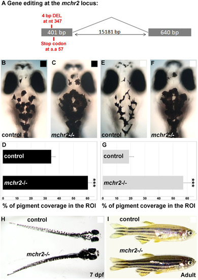
Loss of Mchr2-dependent signaling leads to melanosome dispersion.(A) Schematic representation of the mchr2 locus and the small genomic deletion induced by CRISPR/Cas-9 leading to a premature stop codon in the mchr2 coding sequence. (B, C) Dorsal melanocytes of control (B) or mchr2 homozygous mutant (C) black adapted larvae at 7 dpf. (D) Melanosome coverage was quantified in control or mchr2 homozygous mutant black adapted larvae at 7 dpf in ROI, an area from the posterior of the eyes to the posterior of the hindbrain. (E, F) Dorsal melanocytes of control (E) or mchr2 homozygous mutant (F) white adapted larvae at 7 dpf. (G) Melanosome coverage was quantified in control or mchr2 homozygous mutant white adapted larvae at 7 dpf. (H, I) Larval and adult mchr2 homozygous mutant phenotypes are characterized by melanosome dispersion on a white background, indicating that MCH signaling is required to promote melanosome contraction in melanocytes. Dorsal view with anterior up (B, C, E, F and H). Lateral view (I). Error bars represent s.d. *P<0.05, **P<0.001, ***P<0.0005, determined by t-test, two-tailed.
|