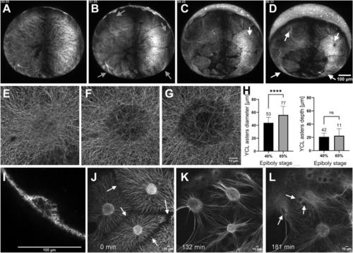
The YCL asters form and evolve throughout epiboly. (A–D) Embryo vegetal view. (A) Before epiboly, the MT network uniformly covers the exposed yolk. (B) At sphere stage, the MT network acquires a new configuration and rearranges into clear domains, with a central high dense MT bundle emanating from each of them (grey arrows). (C) Emerging from those bundles, defined MTs domains start migrating vegetally following a flat to hemisphere transition, leading to the formation of a visible YCL aster (white arrow) with radially oriented MT fibers in the middle of each domain (D) See Video S3. (E–G) High-resolution visualization of an aster formation through CSLM. See also Video S5. (H) After inspection of more than a hundred asters in different embryos we observe that, once formed, asters’ diameter increases with epiboly progression, while their depth remains constant. p-values: ****p < 0.0001, ns means p > 0.05. (I) A cross section of a YCL aster highlights its half-sphere shape. Aster’s dynamics are shown in Video S4 (J) The aster domains remain individualized with defined boundaries (white arrows), with clear opposing MT tips. (K) When the marginal blastoderm approaches the YCL asters (from the top left of the image), the MT network rearranges once again: thicker bundles are formed and reorient in the AV direction, and (L) the cores of the YCL asters actively migrate animalwards, concomitantly with eYSN undergoing epiboly (white arrows), until (L) they disappear underneath the YSL. See Videos S6 and S7.
|
