
FIGURE
Figure 8
Figure 8
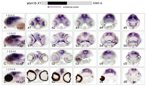
Figure 8. Atxn1b expression domains in cryosectioned zebrafish larvae. Whole-mount in situ hybridization was performed with larvae of the brass and casper line at different developmental stages. Expression domains of atxn1b in whole larva (7 dpf) were detected by antisense probes for the coding region of the transcript (indicated in the upper panel). (A–D) show transverse cryosections (7 µm) of five different regions of the head (left panels). Serial rostral to caudal views of the head region are shown for brass larvae at developmental stages of 4 dpf (a1–a5), 7 dpf (b1–b5), 10 dpf (c1–c5), and for the casper larva at 17 dpf (d1–d5). Abbreviations: cb (cerebellum), dt (dorsal thalamus), ep (epiphysis), ot (optic tectum), pa (pallium), re (retina), rh (rhombencephalon), te (telencephalon), vt (ventral thalamus).
|
Expression Data
| Gene: | |
|---|---|
| Fish: | |
| Anatomical Terms: | |
| Stage Range: | Day 4 to Days 14-20 |
Expression Detail
Antibody Labeling
Phenotype Data
Phenotype Detail
Acknowledgments
This image is the copyrighted work of the attributed author or publisher, and
ZFIN has permission only to display this image to its users.
Additional permissions should be obtained from the applicable author or publisher of the image.
Full text @ Int. J. Mol. Sci.