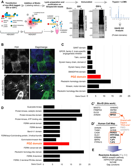
A protein–protein interaction (BioID) screen identifies the PDZ-interactome of GIV-L.A, schematic depicting the key steps in biotin proximity labeling (BioID) studies used to identify the GIV and GIV-L interactomes in HEK293T cells. HEK293T cells were transiently transfected with myc-BirA tagged GIV or GIV-L construct and then treated with free biotin. Equal aliquots of cell lysates were incubated with streptavidin magnetic beads and proteins were eluted by boiling in the presence of excess free biotin. Eluted proteins were analyzed by SDS-PAGE and blotted with AlexaFluor-680-conjugated streptavidin to confirm successful proximity labeling. B, HEK293T cells exogenously expressing myc-BirA-tagged GIV or GIV-L were fixed with methanol prior to staining using anti-myc antibody. Arrows indicate localization onto points of cell–cell contact. Scale bar, 5 μm. C, bar graph summarizing the GIV-L-interacting proteins identified by mass spectrometry and grouped by protein domain using DAVID GO analysis. Top domain categories are shown. C′, list of PDZ domain proteins identified. D, bar graph summarizing GIV's interactome as annotated in the Human Cell map database and also grouped by protein domain using DAVID GO analysis. Top domain categories are shown. Panel D′ lists the PDZ-domain containing proteins reported in the Human Cell Map database.
|
