|
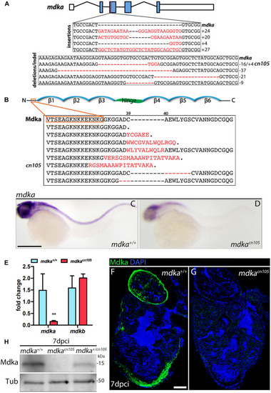
Generation of mdka-KO zebrafish. (A) The mdka locus and DNA mutations introduced by CRISPR/Cas9. The red line indicates the CRISPER target site, red letters indicate insertions, and red hyphens deletions. (B) Mdka domain organization and predicted protein mutations. Red letters denote novel amino acids, red hyphen deletions. β stands for beta-strands and the hinge domain is shown in green. (C,D) Whole-mount ISH (WM-ISH) for mdka in 2-day post-fertilization (dpf) mdka+/+ and mdkacn105 embryos. Scale bar, 200 μm. (E) qPCR analysis of mdka and mdkb in 2 dpf mdka+/+ and mdkacn105 embryos. t-test; **P < 0.01; Mean ± SD. (F,G) Immunofluorescence staining of Mdka in 7 dpci mdka+/+ and mdkacn105 hearts. Scale bar, 100 μm. (H) Western blot analysis for Mdka in 7 dpci mdka+/+, mdkacn105, and mdka+/cn105 hearts. Tub, a-Tubulin; kDa, kilodalton.
|