FIGURE

Figure 4

ID
ZDB-FIG-210519-44
Publication
Kapsokalyvas et al., 2021 - Multiview deconvolution approximation multiphoton microscopy of tissues and zebrafish larvae
Other Figures
All Figure Page
Back to All Figure Page
Figure 4

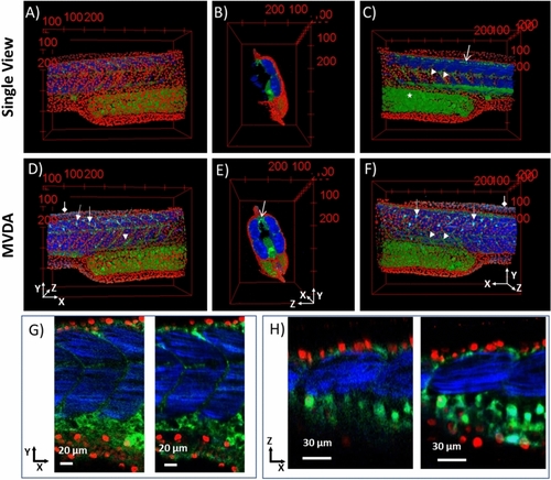
3D images of SV and MVDA. Nuclei in red, autofluorescence and sensory neurons (GFP) in green, muscle fibres and collagen in blue. (A) SV image, (B) SV image rotated 90° to the right. (C) SV rotated 180° compared to (A). (D) MVDA image with the same viewing perspective as A). MVDA image rotated 90° to the right compared to (D). (F) MVDA rotated 180° compared to (D). Arrow heads: myotomes, Diamond arrows collagen in the fins, Open arrows: sensory neurons, Arrows: dendritic like cells. (G) 2D images in the XY plane of SV left and MVDA right. (H) 2D images in the XZ plane of SV left and MVDA right. (Images were created with Fiji26).

Expression Data

Expression Detail
Antibody Labeling
Phenotype Data

Phenotype Detail
Acknowledgments
This image is the copyrighted work of the attributed author or publisher, and ZFIN has permission only to display this image to its users. Additional permissions should be obtained from the applicable author or publisher of the image. Full text @ Sci. Rep.