IMAGE

Figure 4

ID
ZDB-IMAGE-210519-45
Source
Figures for Kapsokalyvas et al., 2021
Image
Figure Caption

Figure 4

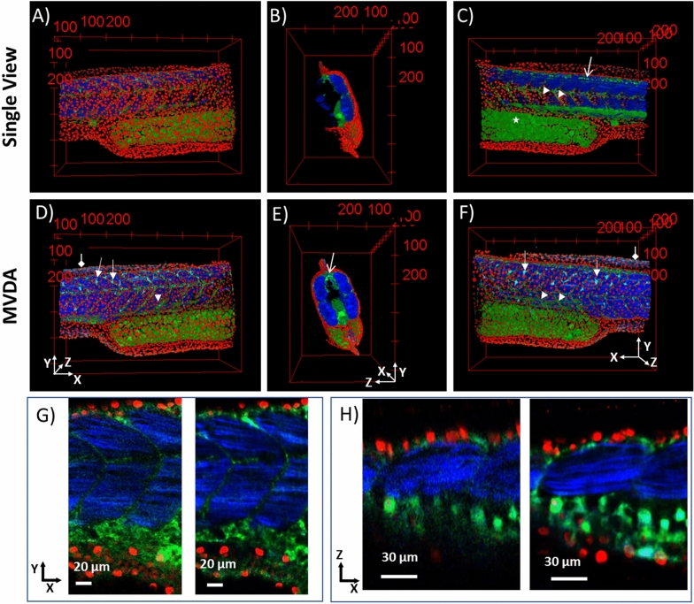
3D images of SV and MVDA. Nuclei in red, autofluorescence and sensory neurons (GFP) in green, muscle fibres and collagen in blue. (A) SV image, (B) SV image rotated 90° to the right. (C) SV rotated 180° compared to (A). (D) MVDA image with the same viewing perspective as A). MVDA image rotated 90° to the right compared to (D). (F) MVDA rotated 180° compared to (D). Arrow heads: myotomes, Diamond arrows collagen in the fins, Open arrows: sensory neurons, Arrows: dendritic like cells. (G) 2D images in the XY plane of SV left and MVDA right. (H) 2D images in the XZ plane of SV left and MVDA right. (Images were created with Fiji26).

Acknowledgments
This image is the copyrighted work of the attributed author or publisher, and ZFIN has permission only to display this image to its users. Additional permissions should be obtained from the applicable author or publisher of the image. Full text @ Sci. Rep.