FIGURE

Figure 1

ID
ZDB-FIG-210519-41
Publication
Kapsokalyvas et al., 2021 - Multiview deconvolution approximation multiphoton microscopy of tissues and zebrafish larvae
Other Figures
All Figure Page
Back to All Figure Page
Figure 1

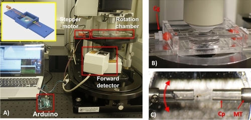
Rotation chamber for Multiview imaging with a two-photon microscope. (A) The rotation chamber mounted on the microscope stage is indicated. The stepper motor is attached to the shaft of the rotation chamber and is controlled by the Arduino board. On the inset of (A) the CAD design of the rotation chamber is shown. (B) The objective is focused on the sample in the rotation chamber. There is ample space for the objective to reach the sample. Below the chamber the condenser lens is positioned. The two cogs responsible for the rotation of the sample are indicated with Cg. (C) An image of the sample when it is ready for imaging. The sample is embedded in the agarose column and is extruded from the capillary allowing the objective to image the sample through the agarose layer. Red arrow indicates rotation direction. MT indicates the metallic tube, Cp indicates the haematocrit glass capillaries.

Expression Data

Expression Detail
Antibody Labeling
Phenotype Data

Phenotype Detail
Acknowledgments
This image is the copyrighted work of the attributed author or publisher, and ZFIN has permission only to display this image to its users. Additional permissions should be obtained from the applicable author or publisher of the image. Full text @ Sci. Rep.