
Fig. 1
- ID
- ZDB-FIG-210507-3
- Publication
- Valera et al., 2021 - A neuronal blueprint for directional mechanosensation in larval zebrafish
- Other Figures
- All Figure Page
- Back to All Figure Page
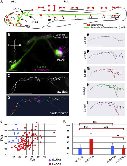
The lateral line in larval zebrafish (A) Schematic representation of a larval zebrafish showing neuromasts (red circles) of the anterior (ALL) and posterior (PLL) lateral line. Posterior neuromasts are named (L for troncal and Ter for terminal), and the planar polarization of their constituent hair cells (horizontal or vertical) is indicated (red arrowheads). Lateralis afferent neurons (LANs) and the lateralis central lintel (LCL) are shown in green. (B) Confocal image of a HGn39D transgenic larva (green) injected with Sill:mCherry (red), showing the neuronal soma of a posterior LAN (pLAN) within the posterior ganglion (PLLG) and its position within the LCL. The anterior ganglion (ALLG) is also shown. (C) Structure of the central projection of an individually marked LAN. (D) Skeletonization of the same projection shown in (C) with terminal varicosities marked with colored dots. (E–I) Skeletonized central arbors of the same neuron over consecutive days from 2.5 to 6.5 days-post fertilization (dpf). In all panels, anterior is left and dorsal is up. (J) aLANs and pLANs (respectively, blue diamonds and red squares) constitute distinguishable morphological classes according to the number of anterior and posterior varicosities (respectively AVs and PVs). (K) Bar chart showing significant differences between aLANs (blue) and pLANs (red) for AVs and PVs. aLANs have similar number of AVs and PVs along the length of the central projection. Scale bars are 50 μm. See also Figure S1. |