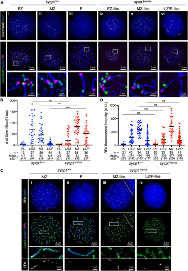
DSB localization in sycp1 mutant spermatocytes. (A) costaining of Dmc1/Rad51, telomeres and Sycp3 in sycp1+/+ (i to iii) and sycp1isa/isa (iv to vi) spermatocytes. (B) quantification of Dmc1/Rad51 focus numbers in sycp1+/+ and sycp1isa/isa spermatocytes. Black bars indicate means. Statistical significance was examined by a two-tailed Mann-Whitney test (**P < 0.01, ***P < 0.001, ****P < 0.0001; exact P value). (C) costaining of RPA, Sycp1 and Sycp3 in sycp1+/+ (i and ii) and sycp1isa/isa (iii and iv) spermatocytes. Regions marked as a white rectangle in the middle panels are shown at a higher magnification at the bottom. The white line on (iii) indicates a border with another nucleus on the top right. (D) quantification of RPA signal intensity in sycp1+/+ and sycp1isa/isa spermatocytes. Black bars indicate means. Statistical significance was examined by two-tailed Mann-Whitney test (ns, not significant; exact P value). Since RPA is observed as short stretches rather than discrete foci, signal intensity rather than the number of foci in a nucleus was quantified here. Quantification of RPA focus numbers is shown in Supplementary Figure 5.
|
