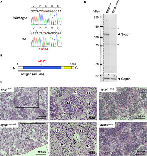
A premature stop mutation of the sycp1 gene in the isa mutant line. (A) Genomic sequences of a part of exon 15 in the sycp1 gene. DNA sequences and peaks obtained from a wild-type and an isa mutant fish are shown with corresponding amino acids (above in 1-letter abbreviations). Positions of the isa mutation (base 1225 of the sycp1 coding sequence) are shown in red letters. (B) A schematic presentation of the Sycp1 protein sequence. The isa mutation site is indicated with an arrow. The antigen region (amino acids 1 to 408) of the Sycp1 antibody used in this study is shown below as a black bar. N: N-terminus, C: C-terminus. Numbers indicate corresponding amino acid positions. (C) Western blotting of the Sycp1 protein. Testis protein extracts of wild-type (sycp1+/+) and isa homozygote mutant (sycp1isa/isa) adult fish were blotted with anti-Sycp1 antibody and anti-Gapdh antibody as an internal control. The left side of the broken line is a part labeled with a molecular ladder on the same membrane. The predicted molecular sizes of full-length Sycp1 and Gapdh proteins are 116 kDa and 35.8 kDa, respectively. Non-specific bands were marked with asterisks (*). (D) HE-stained sections of sycp1+/+, sycp1isa/del5, sycp1isa/+ and sycp1del5/+ testes. All samples were prepared from siblings at 4 mpf (months post fertilization). Representative results of 3 individual fish are shown for each genotype. Magnified images are shown for sycp1+/+ and sycp1isa/del5 sections. Sc: spermatocytes, St: spermatids, Sz: spermatozoa. In the sycp1isa/del5 section, lumens with no spermatozoa (asterisks) and spermatocytes with irregular nuclei (inside broken lines) were observed.
|
