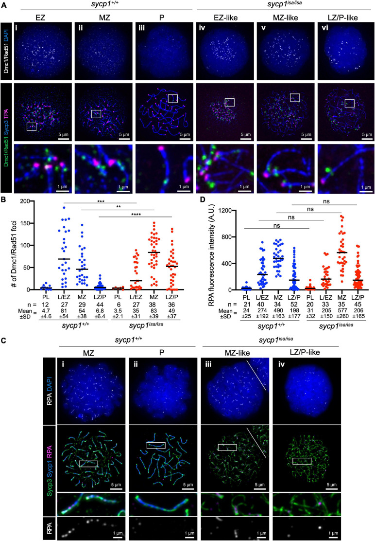
IMAGE

FIGURE 6

ID
ZDB-IMAGE-210414-26
Source
Figures for Imai et al., 2021
Image
Figure Caption

FIGURE 6

DSB localization in sycp1 mutant spermatocytes. (A) costaining of Dmc1/Rad51, telomeres and Sycp3 in sycp1+/+ (i to iii) and sycp1isa/isa (iv to vi) spermatocytes. (B) quantification of Dmc1/Rad51 focus numbers in sycp1+/+ and sycp1isa/isa spermatocytes. Black bars indicate means. Statistical significance was examined by a two-tailed Mann-Whitney test (**P < 0.01, ***P < 0.001, ****P < 0.0001; exact P value). (C) costaining of RPA, Sycp1 and Sycp3 in sycp1+/+ (i and ii) and sycp1isa/isa (iii and iv) spermatocytes. Regions marked as a white rectangle in the middle panels are shown at a higher magnification at the bottom. The white line on (iii) indicates a border with another nucleus on the top right. (D) quantification of RPA signal intensity in sycp1+/+ and sycp1isa/isa spermatocytes. Black bars indicate means. Statistical significance was examined by two-tailed Mann-Whitney test (ns, not significant; exact P value). Since RPA is observed as short stretches rather than discrete foci, signal intensity rather than the number of foci in a nucleus was quantified here. Quantification of RPA focus numbers is shown in Supplementary Figure 5.

Acknowledgments
This image is the copyrighted work of the attributed author or publisher, and ZFIN has permission only to display this image to its users. Additional permissions should be obtained from the applicable author or publisher of the image. Full text @ Front Cell Dev Biol