
FIGURE
Figure 4—figure supplement 1.
- ID
- ZDB-FIG-210319-16
- Publication
- Vinayagam et al., 2020 - Structural basis of TRPC4 regulation by calmodulin and pharmacological agents
- Other Figures
-
- Figure 1.
- Figure 2
- Figure 2—figure supplement 1.
- Figure 2—figure supplement 2.
- Figure 2—figure supplement 3.
- Figure 2—figure supplement 4.
- Figure 2—figure supplement 5.
- Figure 3
- Figure 3—figure supplement 1.
- Figure 4
- Figure 4—figure supplement 1.
- Figure 4—figure supplement 2.
- Figure 5
- Figure 5—figure supplement 1.
- Figure 5—figure supplement 2.
- Figure 5—figure supplement 3.
- Figure 5—figure supplement 4.
- Figure 5—figure supplement 5.
- Figure 5—figure supplement 6.
- Figure 6.
- Figure 7.
- All Figure Page
- Back to All Figure Page
Figure 4—figure supplement 1.
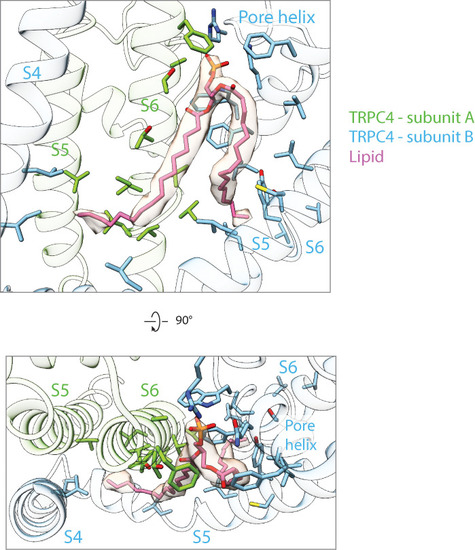
Phosphatidic acid (pink) that binds at the interface between two subunits near the pore region is shown in stick representation along with the corresponding density. The interacting residues from the S4, S5 and S6 helices are also shown in stick representation. The protein residues from different protomers are colored differently. The helices are shown in cartoon representation with high transparency. |
Expression Data
Expression Detail
Antibody Labeling
Phenotype Data
Phenotype Detail
Acknowledgments
This image is the copyrighted work of the attributed author or publisher, and
ZFIN has permission only to display this image to its users.
Additional permissions should be obtained from the applicable author or publisher of the image.
Full text @ Elife