|
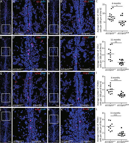
<italic>Δ113p53</italic> promotes cell proliferation in the ventricular zone of the telencephalon.A–F The Δ113p53+/+ (A, D) and Δ113p53M/M (B, E) zebrafish telencephalons were labeled with EDU in red at 6- and 11-months old, as indicated. G–L Cryosections of Δ113p53+/+ (G, J) and Δ113p53M/M (H, K) zebrafish telencephalons were immunostained by anti-PCNA (in red) at 6- and 11-months old, as indicated. The nuclei were stained with DAPI (in blue). An average number of EDU+ cells or PCNA+ cells per 100 μm of the ventricular zone (VZ) in one section from the middle region of each telencephalon was presented in C (6-months old) and F (11-months old) or I (6-months old) and L (11-months old), respectively. Framed areas in A, B, D, E, G, H, J, K were magnified in A’, B’, D’, E’, G’, H’, J’, K’, respectively. Scale bar in A, B, D, E, G, H, J, K, 50 μm; scale bar in A’, B’, D’, E’, G’, H’, J’, K’, 10 μm. Each dot represents the average number of EDU+ cells or PCNA+ cells per 100 μm of VZ in one section. About three to five sections were chosen from the middle region of each telencephalon and at least three telencephalons were sampled in each group. Statistical analysis was performed on relevant data using the Student’s two-tailed t test. *P < 0.05, **P < 0.01, ***P < 0.001.
|