Fig. 2
- ID
- ZDB-FIG-210216-70
- Publication
- Rieckhoff et al., 2020 - Spindle Scaling Is Governed by Cell Boundary Regulation of Microtubule Nucleation
- Other Figures
- All Figure Page
- Back to All Figure Page
|
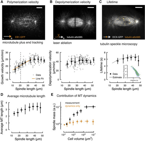
Figure 2. Microtubule Dynamics Are Insufficient to Account for Spindle Scaling (A) Quantification of microtubule polymerization in spindles of different sizes during early zebrafish development shows two regimes (see also Figure S2 and Video S3). Microtubule growth velocity scales linearly with spindle length (aster-aster) in spindles smaller than 35 μm. In larger spindles, microtubule polymerization velocity remains constant. Individual and binned measurements are shown in gray (n = 196) and black, respectively (mean ± SD, bin size 5 μm). Gray dotted line represents the estimated transition to zygotic genome activation (ZGA) corresponding to spindles on the left side of the line. Scale bar, 10 μm. (B) Laser ablation reveals that microtubule depolymerization velocity is independent of spindle length (see also Figure S3 and Video S4; n = 202, mean ± SD, bin size 5 μm). Scale bar, 10 μm. (C) Tubulin speckle microscopy reveals that microtubule lifetimes are constant in spindles of different sizes (see also Figure S4 and Video S5). Solid gray line is the estimate derived from changes in the microtubule polymerization velocity observed during scaling (STAR Methods). Inset shows the measured speckle lifetime distributions color-coded by spindle length (n = 47, mean ± SD). Scale bar, 10 μm. (D) Microtubule (MT) length correlates with spindle size in spindles smaller than 35 μm but remains constant in larger spindles. Average microtubule length obtained from the measured microtubule dynamics (STAR Methods). Error bars are obtained from error propagation of the measurements of microtubule dynamics. (E) Modulation of microtubule dynamics is not sufficient to account for spindle scaling. The estimated scaling of spindle mass from microtubule dynamics alone (in orange) is significantly lower than the measured spindle mass (in black). |

