
Fig. 1
- ID
- ZDB-FIG-210216-69
- Publication
- Rieckhoff et al., 2020 - Spindle Scaling Is Governed by Cell Boundary Regulation of Microtubule Nucleation
- Other Figures
- All Figure Page
- Back to All Figure Page
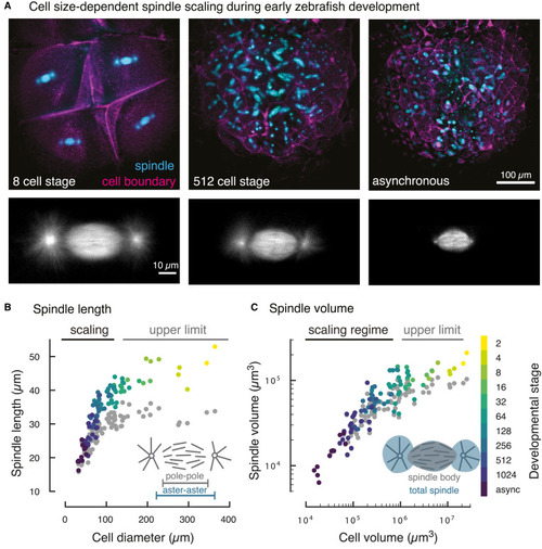
Figure 1. Cell Size-Dependent Spindle Scaling during Early Zebrafish Development (A) 3D light-sheet microscopy of spindles and cell boundaries in zebrafish embryos. Maximum intensity projections of a fluorescently labeled zebrafish embryo at selected developmental stages (see Video S1). (B) Spindle scaling in early zebrafish embryos covers two regimes. In large cells, spindle length remains constant (“upper limit”). Below a critical cell diameter (dcell = 125 μm), spindle length scales linearly with cell diameter. Each dot denotes an individual measurement of aster-to-aster spindle length (color-coded by developmental stage) or pole-to-pole spindle length (in gray, 96 analyzed spindles and cells in four different embryos). (C) 3D segmentation of spindles and cells (see Figure S1) yields the scaling relationship between spindle volume and cell volume. The scaling of the total spindle volume (including asters, color-coded by cell stage) and the spindle body volume (excluding asters, gray) is nearly indistinguishable. |