FIGURE

Figure 11

ID
ZDB-FIG-201229-87
Publication
Candiani et al., 2020 - Alexander Disease Modeling in Zebrafish: An In Vivo System Suitable to Perform Drug Screening
Other Figures
All Figure Page
Back to All Figure Page
Figure 11

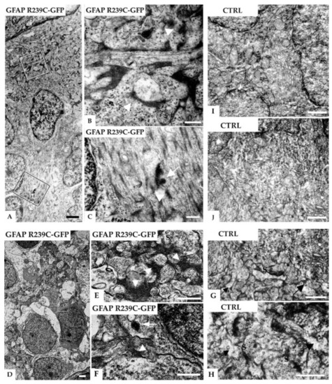
Electron micrographs of radial glial cells in the subventricular and neuropil region in the telencephalon of a 5 dpf zebrafish injected with GFAP-R239C-GFP plasmid. (A) A glial cell contacts the ventricular brain surface covered by the tela choroidea (top) and shows many electron-dense profiles. In (B,C), higher magnifications of the marked areas in (A) reveal that the dark profiles are associated with filaments (arrowheads) and sometimes surrounded by a membrane (arrows), the latter consistent with lysosomal packaging. In another example of the neuropil region shown in (D), at higher magnifications of the marked areas depicted in (E,F), glial processes containing electron-dense fibrillar material can be seen (arrowheads). The arrow points at a membrane-bound vesicle containing clustered electron-dense material, featuring a lysosome. In (GJ), electron microscopic analyses on control wildtype embryos of similar regions are shown, i.e., subventricular glial processes in (G,H) and neuropil region in the telencephalon in (I,J). No such electron-dense material in glial processes, identified by their irregular shape in the synaptic regions (arrows), was detected in the control tissue. Note that the dark bands are postsynaptic densities (S: presynaptic terminals). Scale bars: (A,D) 1 µm; (B,C) 250 nm; (EJ) 500 nm.

Expression Data

Expression Detail
Antibody Labeling
Phenotype Data

Phenotype Detail
Acknowledgments
This image is the copyrighted work of the attributed author or publisher, and ZFIN has permission only to display this image to its users. Additional permissions should be obtained from the applicable author or publisher of the image. Full text @ Genes (Basel)