
FIGURE
Figure 4
- ID
- ZDB-FIG-201229-80
- Publication
- Candiani et al., 2020 - Alexander Disease Modeling in Zebrafish: An In Vivo System Suitable to Perform Drug Screening
- Other Figures
- All Figure Page
- Back to All Figure Page
Figure 4
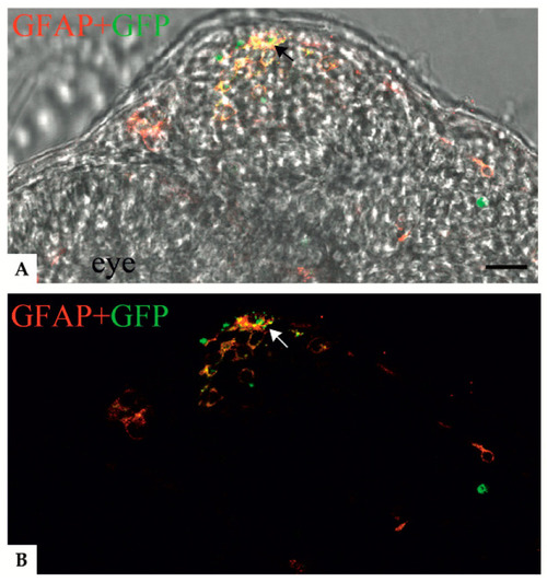
Confocal images showing colocalization between anti-GFAP signal and the GFAP R239C-GFP aggregates in zebrafish transverse sections of 24 hpf embryos. ( |
Expression Data
Expression Detail
Antibody Labeling
Phenotype Data
Phenotype Detail
Acknowledgments
This image is the copyrighted work of the attributed author or publisher, and
ZFIN has permission only to display this image to its users.
Additional permissions should be obtained from the applicable author or publisher of the image.
Full text @ Genes (Basel)