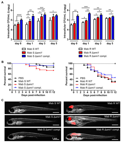
Assessment of Δami1Mab in human macrophage and zebrafish larvae. (A) M. abscessus-infected THP-1 macrophages with amikacin maintained at 50 μg/mL in the RPMI medium were lysed from day 0 to day 5 for CFU enumeration. Bacteria were plated at day 0 (2–4 h phagocytosis followed by a 2 h amikacin treatment at 250 μg/mL) day 1, 3 and day 5. Histograms and error bars represent means and standard deviations calculated from three independent experiments. For statistical analysis, the unpaired t-test with Welch’s correction was applied. *, **, ***, and **** stand for p < 0.1, p < 0.01, p < 0.001, and p < 0.0001, respectively. Left and right panels are for M. abscessus S and R variants, respectively. The M. abscessus S and R wild-type strains used as controls were not transformed with pMV306. (B) Survival curves of zebrafish embryos infected with the different M. abscessus strains. The data shown are corresponding to a pool of data from three independent experiments. For each experiment, 20 embryos were injected with 200–300 CFUs, respectively. Left and right panels are for M. abscessus S and R variants, respectively. The M. abscessus S and R wild-type strains used as controls were transformed with pTEC27 but not transformed with pMV306. (C) Representative images of infected zebrafish embryos at 5 days post-infection. Scale bar represents 1 mm. Infection foci are displayed in red.
|
