
FIGURE
Figure 6
- ID
- ZDB-FIG-201130-72
- Publication
- Küssau et al., 2020 - Functional Characterization of the N-Acetylmuramyl-l-Alanine Amidase, Ami1, from Mycobacterium abscessus
- Other Figures
- All Figure Page
- Back to All Figure Page
Figure 6
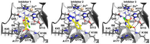
In silico docking of the three inhibitors. The figure depicts the best docking poses of the three compounds. All three inhibitors appear able to bind in the Ami1Mab active site with similar binding energies. Inhibitors are displayed in stick representation. Nitrogen atoms are in blue, oxygen in red, hydrogen in white and carbon atoms in yellow for the ligand and grey for the protein. |
Expression Data
Expression Detail
Antibody Labeling
Phenotype Data
Phenotype Detail
Acknowledgments
This image is the copyrighted work of the attributed author or publisher, and
ZFIN has permission only to display this image to its users.
Additional permissions should be obtained from the applicable author or publisher of the image.
Full text @ Cells