FIGURE

FIGURE 3

ID
ZDB-FIG-201003-22
Publication
Sundaramurthi et al., 2020 - Selective Histone Deacetylase 6 Inhibitors Restore Cone Photoreceptor Vision or Outer Segment Morphology in Zebrafish and Mouse Models of Retinal Blindness
Other Figures
All Figure Page
Back to All Figure Page
FIGURE 3

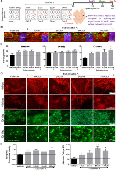
TubA preserves cone cells in rd10 mice retinal explants. (A) Schematic diagram outlining the drug treatment protocol in retinal explants from homozygous rd10/rd10 mice (B6.CXBI–Pde6brd10/J) at postnatal day 14 (P14). (B) Retinal section fluorescent images of explants treated with increasing concentrations of TubA or vehicle control, labeled for rhodopsin (rods) and cone arrestin (cones). The total number of nuclei in the outer nuclear layer (ONL) did not change significantly across the TubA treatment group. Likewise, the number of rhodopsin positive outer segments did not significantly change. A dose-dependent increase in the number of cone arrestin positive outer segments was observed, with a significant increase following 100 μM TubA treatment. (C) In wholemount preparations, no significant increase in fluorescence intensity of rhodopsin positive cells, was observed following TubA treatment. A significant increase in the number of cells with cone arrestin positive outer segments was identified at 50 μM and 100 μM TubA treatment. Statistical analysis was performed using One-way ANOVA with Dunnett’s multiple comparisons. N = 4 per treatment group.

Expression Data

Expression Detail
Antibody Labeling
Phenotype Data

Phenotype Detail
Acknowledgments
This image is the copyrighted work of the attributed author or publisher, and ZFIN has permission only to display this image to its users. Additional permissions should be obtained from the applicable author or publisher of the image. Full text @ Front Cell Dev Biol