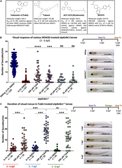
Selective HDAC6i significantly restores visual function in atp6v0e1–/–, a zebrafish model of retinal blindness. (A) Table describing the physiochemical properties of the four HDAC6i candidates selected for present study. Chemical structures were drawn using Chemspider (http://www.chemspider. com/StructureSearch.aspx). (B)atp6v0e1–/– mutants were treated at maximum tolerated concentration (refer to Supplementary Figures S1, S2A) of each HDAC6i, from 3–6 days post fertilization (dpf). Visual function was markedly improved upon treatment with 100 μM TubA, 10 μM Tubacin, 10 μM ACY-1215 or 25 μM NF2373 compared to vehicle-control (0.1% DMSO) treated larvae. Panel on right shows representative larval wholemount images. Statistical analysis was performed using One-way ANOVA with Dunnett’s multiple comparisons, where ∗∗∗p-value = 0.0008 and **** means a p-value of ≤ 0.0001. N = 3 and n = 12 per treatment group. (C) The duration of action of TubA was determined, whereby atp6v0e1–/– larvae were treated from 3 to 8 dpf, and visual function measured at 6, 7 or 8 dpf (refer to Supplementary Figure S2B for visual response in siblings). A significant increase in visual response was recorded at 7 and 8 dpf, respectively. Representative larval images presented in the right panel. Student’s T test was used for statistical analysis, where ∗∗∗ means a p-value of ≤ 0.0001. N = 3 and n = 12 per treatment group.
|
