FIGURE

FIGURE 2

ID
ZDB-FIG-201003-21
Publication
Sundaramurthi et al., 2020 - Selective Histone Deacetylase 6 Inhibitors Restore Cone Photoreceptor Vision or Outer Segment Morphology in Zebrafish and Mouse Models of Retinal Blindness
Other Figures
All Figure Page
Back to All Figure Page
FIGURE 2

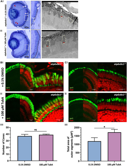
TubA treatment improved retinal morphology in atp6v0e1–/– larvae. (Ai) LM images revealed large vacuoles (pink arrows) in the RPE layer, pyknotic nuclei (red arrows) in the CMZ and shortened photoreceptor outer segments in vehicle-control treated atp6v0e1–/– mutants. (Aii) The photoreceptor outer segments were observed to be more elongated, more prominent, and better organized in TubA-treated atp6v0e1–/– mutants compared to vehicle-control treated atp6v0e1–/– mutants (Supplementary Figure S3). (B,C) Confocal images of immuno-stained vehicle-control or TubA-treated atp6v0e1–/– mutant retina with cone (zpr-1) photoreceptor and rod and cone outer segment (zpr-3) specific markers. (Bi,Ci) Panels present aberrant cone and rod outer segment morphology in vehicle-control treated atp6v0e1–/– mutants. (Bii,Cii) TubA treatment improved photoreceptor structural integrity and organization in atp6v0e1–/– mutants. (Biii,Ciii) Graphical presentation of the number of cone cells (zpr-1 staining) and total area occupied by photoreceptor outer segments (zpr-3 staining), respectively (refer to Supplementary Figure S4 for analysis of retinal histology in siblings; 3 sections per sample was analyzed, N = 3). Student’s unpaired T-test was used for statistical analysis.

Expression Data

Expression Detail
Antibody Labeling
Phenotype Data
Fish:
Condition:
Observed In:
Stage: Day 6

Phenotype Detail
Acknowledgments
This image is the copyrighted work of the attributed author or publisher, and ZFIN has permission only to display this image to its users. Additional permissions should be obtained from the applicable author or publisher of the image. Full text @ Front Cell Dev Biol