|
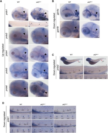
Altered expression of oxidative stress markers in ak2hg14 embryos. Expression analysis of oxidative stress markers in ak2hg14 and control sibling (WT) embryos at 4 or 5 dpf. (A) Upregulated expression of several prdx genes in the otic vesicle and PLL at 4 dpf. Bottom panel for prdx1 marker: the black arrows indicate ectopic expression of the marker in PLL neuromasts of the corresponding embryos. (B) Downregulated expression of gpx4a and gpx4b markers at 4 dpf. (C) Specific downregulation of prdx2 marker at 5 dpf in PLL neuromasts and caudal hematopoietic tissue (red arrowhead, CHT). Scale bars: 200 µm. (D) Comparison of gstp1 expression in the PLL neuromasts in control embryos, ak2hg14 null and ak2hg16 hypomorphic mutants at 4 dpf. Two different embryos per genotype are shown. Lateral views with anterior to the left. Black arrowheads indicate the otic vesicle. Scale bars: 100 µm.
|